3.2 Information Processing in Neurons
Our thoughts, feelings, and actions depend on neural communication, but how does it happen? The communication of information within and between neurons proceeds in two stages. First, information has to travel inside the neuron via an electrical signal that travels from the dendrite to the cell body to the axon—
Electric Signaling: Conducting Information within a Neuron
The neuron’s cell membrane has small pores that act as channels to allow small electrically charged molecules, called ions, to flow in and out of the cell. It is this flow of ions across the neuron’s cell membrane that creates the conduction of an electric signal within the neuron. How does it happen?
The Resting Potential: The Origin of the Neuron’s Electrical Properties
Neurons have a natural electric charge called the resting potential, the difference in electric charge between the inside and outside of a neuron’s cell membrane (Kandel, 2000). The resting potential arises from the difference in concentrations of ions inside and outside the neuron’s cell membrane (see FIGURE 3.3a). Ions can carry a positive (+) or a negative (-) charge. In the resting state, there is a high concentration of a positively charged ion, potassium (K+), as well as negatively charged protein ions (A–), inside the neuron’s cell membrane compared to outside it. By contrast, there is a high concentration of positively charged sodium ions (Na+) and negatively charged chloride ions (Cl–) outside the neuron’s cell membrane.
resting potential
The difference in electric charge between the inside and outside of a neuron’s cell membrane.
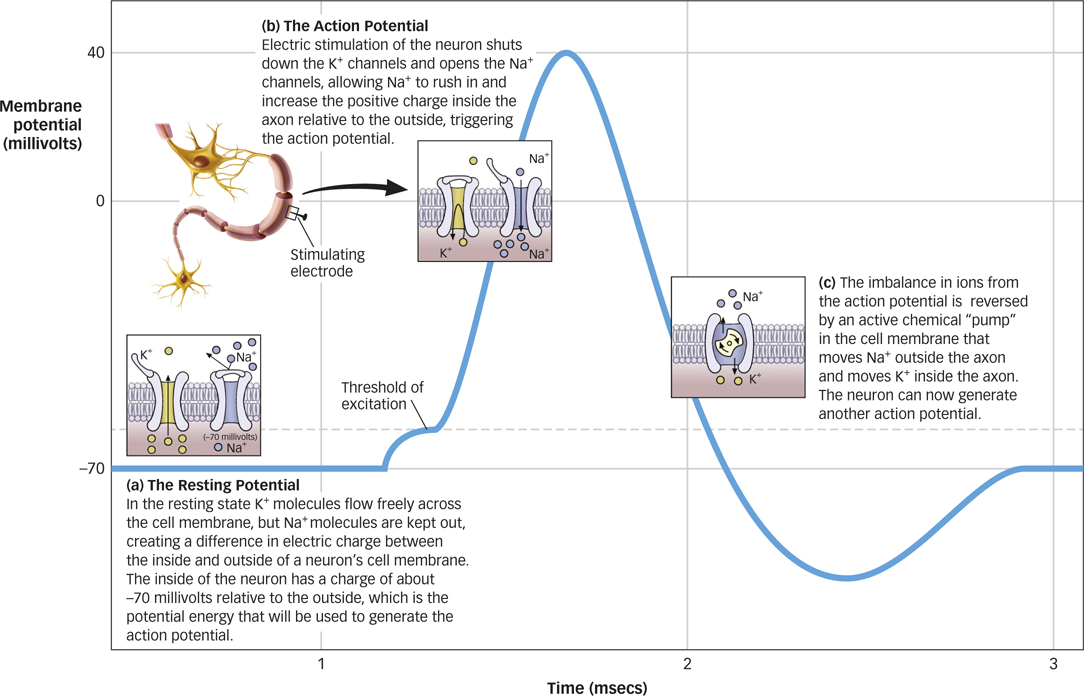
The concentration of K+ inside and outside a neuron is controlled by channels in the cell membrane that allow K+ molecules to flow in and out of the neuron. In the resting state, the channels that allow K+ molecules to flow freely across the cell membrane are open, while channels that allow the flow of Na+ and the other ions are generally closed. Because of the naturally higher concentration of K+ molecules inside the neuron, some K+ molecules move out of the neuron through the open channels, leaving the inside of the neuron with a charge of about -70 millivolts relative to the outside. Like the Hoover Dam that holds back the Colorado River until the floodgates are released, resting potential is potential energy, because it creates the environment for a possible electrical impulse.
What difference between the inside and outside of the neuron’s cell membrane creates the resting potential?
60
The Action Potential: Sending Signals across the Neuron
The neuron maintains its resting potential most of the time. However, in the 1930s, biologists Alan Hodgkin and Andrew Huxley noticed that they could produce a signal by stimulating the axon with a brief electric shock, which resulted in the conduction of an electric impulse down the length of the axon (Hausser, 2000; Hodgkin & Huxley, 1939). This electric impulse is called an action potential, an electric signal that is conducted along the length of a neuron’s axon to a synapse.
action potential
An electric signal that is conducted along a neuron’s axon to a synapse.
The action potential occurs only when the electric shock reaches a certain level, or threshold. The action potential is all or none: Electric stimulation below the threshold fails to produce an action potential, whereas electric stimulation at or above the threshold always produces the action potential. The action potential always occurs with exactly the same characteristics and at the same magnitude regardless of whether the stimulus is at or above the threshold.
Why is an action potential an all-
The action potential occurs when there is a change in the state of the axon’s membrane channels. Remember, during the resting potential, the channels that allow K+ to flow out are open, resulting in a net negative charge (–70 millivolts) relative to the outside. However, during an action potential, these channels briefly shut down, and channels that allow the flow of positively charged sodium ions (Na+) are opened (see FIGURE 3.3b). We’ve seen already that Na+ is typically much more concentrated outside the axon than inside. When the channels open, those positively charged ions (Na+) flow inside, increasing the positive charge inside the axon relative to the outside. This flow of Na+ into the axon pushes the action potential from negative (–70 millivolts) to positive (+40 millivolts).
61
After the action potential reaches its maximum, the membrane channels return to their original state, and K+ flows out until the axon returns to its resting potential. This leaves a lot of extra Na+ ions inside the axon and a lot of extra K+ ions outside the axon. During this period when the ions are imbalanced, the neuron cannot initiate another action potential, so it is said to be in a refractory period, the time following an action potential during which a new action potential cannot be initiated. The imbalance in ions is eventually reversed by an active chemical “pump” in the cell membrane that moves Na+ outside the axon and moves K+ inside the axon (the pump does not operate during the action potential; see FIGURE 3.3c).
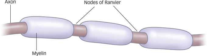
So far, we’ve described how the action potential occurs at one point in the neuron. But how does this electric charge move down the axon? It’s a domino effect. When an action potential is generated at the beginning of the axon, it spreads a short distance, which generates an action potential at a nearby location on the axon, and so on, thus conducting the charge down the length of the axon.
The myelin sheath facilitates the conduction of the action potential. Myelin doesn’t cover the entire axon; rather, it clumps around the axon with little break points between clumps, looking kind of like sausage links. These break points are called the nodes of Ranvier, after French pathologist Louis-
Chemical Signaling: Transmission between Neurons
When the action potential reaches the end of an axon, you might think that the action potential stops there. After all, the synaptic space between neurons means that the axon of one neuron and the neighboring neuron’s dendrites do not actually touch one another. However, the electric charge of the action potential takes a form that can cross the relatively small synaptic gap by relying on a bit of chemistry.
Axons usually end in terminal buttons, knob-
terminal buttons
Knoblike structures that branch out from an axon.
neurotransmitters
Chemicals that transmit information across the synapse to a receiving neuron’s dendrites.
receptors
Parts of the cell membrane that receive the neurotransmitter and initiate or prevent a new electric signal.
How does a neuron communicate with another neuron?
62
The action potential travels down the length of the axon of the sending neuron, or presynaptic neuron, to the terminal buttons, where it stimulates the release of neurotransmitters from vesicles into the synapse. These neurotransmitters float across the synapse and bind to receptor sites on a nearby dendrite of the receiving neuron, or postsynaptic neuron. A new electric signal is initiated in that neuron, which may in turn generate an action potential in that neuron. This electrochemical action, called synaptic transmission (see FIGURE 3.5), allows neurons to communicate with one another.
Neurotransmitters and receptor sites act like a lock-
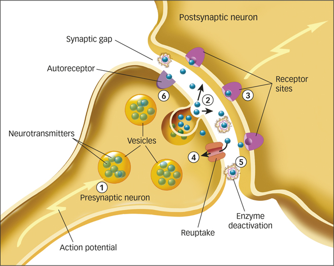
What happens to the neurotransmitters left in the synapse after the chemical message is relayed to the postsynaptic neuron? Something must make neurotransmitters stop acting on neurons; otherwise, there’d be no end to the signals that they send. Neurotransmitters leave the synapse through three processes (see FIGURE 3.5). First, neurotransmitters can be reabsorbed by the terminal buttons of the presynaptic neuron’s axon, a process called reuptake. Second, neurotransmitters can be destroyed by enzymes in the synapse, in a process called enzyme deactivation. Third, neurotransmitters can bind to receptor sites called autoreceptors on the presynaptic neuron. Autoreceptors detect how much of a neurotransmitter has been released into a synapse and signal the presynaptic neuron to stop releasing the neurotransmitter when an excess is present.
Types and Functions of Neurotransmitters
You might wonder how many types of neurotransmitters are floating across synapses in your brain right now. Today, we know that some 60 chemicals play a role in transmitting information throughout the brain and body and differentially affect thought, feeling, and behavior, but a few major classes seem particularly important. We’ll summarize those here, and you’ll meet some of these neurotransmitters again in later chapters.

- Acetylcholine (ACh) is a neurotransmitter involved in a number of functions, including voluntary motor control. Acetylcholine is found in neurons of the brain and in the synapses where axons connect to muscles and body organs, such as the heart. Acetylcholine contributes to the regulation of attention, learning, sleeping, dreaming, and memory (Gais & Born, 2004; Hasselmo, 2006; Wrenn et al., 2006). Alzheimer’s disease, a medical condition involving severe memory impairments (Salmon & Bondi, 2009), is associated with the deterioration of ACh-
producing neurons. - Dopamine is a neurotransmitter that regulates motor behavior, motivation, pleasure, and emotional arousal. Because of its role in associating actions with rewards, dopamine plays a role in drug addiction (Baler & Volkow, 2006). High levels of dopamine have been linked to schizophrenia (Winterer & Weinberger, 2004), whereas low levels have been linked to Parkinson’s disease.
- Glutamate is the major excitatory neurotransmitter in the brain, meaning that it enhances the transmission of information between neurons. GABA (gamma-
aminobutyric acid), in contrast, is the primary inhibitory neurotransmitter in the brain, meaning that it tends to stop the firing of neurons. Too much glutamate, or too little GABA, can cause neurons to become overactive, causing seizures. - Two related neurotransmitters influence mood and arousal: norepinephrine and serotonin. Norepinephrine is involved in vigilance, or a heightened awareness of dangers in the environment (Ressler & Nemeroff, 1999). Serotonin is involved in the regulation of sleep and wakefulness, eating, and aggressive behavior (Dayan & Huys, 2009; Kroeze & Roth, 1998). Because both neurotransmitters affect mood and arousal, low levels of each have been implicated in mood disorders (Tamminga et al., 2002).
- Endorphins are chemicals that act within the pain pathways and emotion centers of the brain (Keefe et al., 2001). Endorphins help dull the experience of pain and elevate moods. The “runner’s high” experienced by many athletes as they push their bodies to painful limits of endurance can be explained by the release of endorphins in the brain (Boecker et al., 2008).
63
How do neurotransmitters create the feeling of runner’s high?
How Drugs Mimic Neurotransmitters
Each of these neurotransmitters affects thought, feeling, and behavior in different ways, so normal functioning involves a delicate balance of each. Even a slight imbalance—
Many drugs that affect the nervous system operate by increasing, interfering with, or mimicking the manufacture or function of neurotransmitters (Cooper, Bloom, & Roth, 2003; Sarter, 2006). Agonists are drugs that increase the action of a neurotransmitter. Antagonists are drugs that block the function of a neurotransmitter (see FIGURE 3.6).
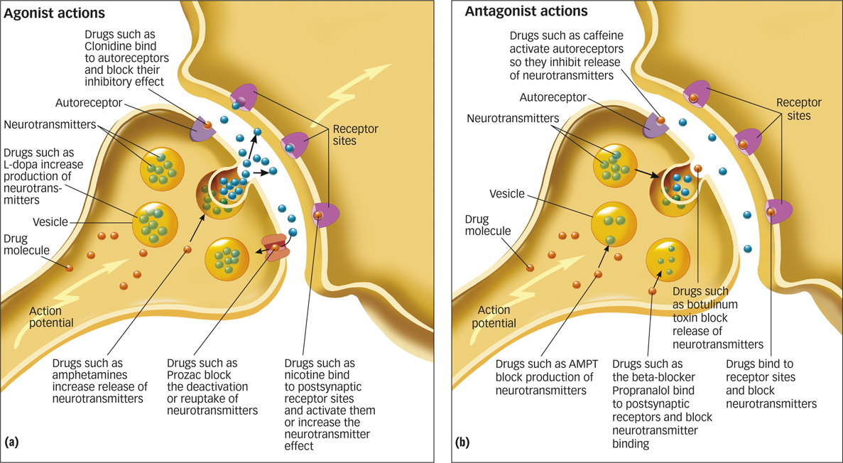

agonists
Drugs that increase the action of a neurotransmitter.
antagonists
Drugs that block the function of a neurotransmitter.
For example, the drug L-
How does L-
As another example, amphetamine is a popular drug that stimulates the release of norepinephrine and dopamine. In addition, both amphetamine and cocaine prevent the reuptake of norepinephrine and dopamine. The combination of increased release of norepinephrine and dopamine and the prevention of their reuptake floods the synapse with those neurotransmitters, resulting in increased activation of their receptors. Both of these drugs, therefore, are strong agonists, although the psychological effects of the two drugs differ somewhat because of subtle distinctions in where and how they act on the brain. Norepinephrine and dopamine play a critical role in mood control, such that increases in either neurotransmitter result in euphoria, wakefulness, and a burst of energy. However, norepinephrine also increases heart rate. An overdose of amphetamine or cocaine can cause the heart to contract so rapidly that heartbeats do not last long enough to pump blood effectively, leading to fainting and sometimes death.
64
Other drugs that mimic neurotransmitters, such as antianxiety and antidepression drugs, will be discussed later, in the Treatment of Psychological Disorders chapter.
SUMMARY QUIZ [3.2]
Question 3.5
| 1. | An electric signal that is conducted along the length of a neuron’s axon to the synapse is called |
- a resting potential.
- an action potential.
- a node of Ranvier.
- an ion.
b.
Question 3.6
| 2. | The chemicals that transmit information across the synapse to a receiving neuron’s dendrites are called |
- vesicles.
- terminal buttons.
- postsynaptic neurons.
- neurotransmitters.
d.
65