4.4 Audition: More Than Meets the Ear
Vision is based on the spatial pattern of light waves on the retina. The sense of hearing, by contrast, is all about sound waves: changes in air pressure unfolding over time. Plenty of things produce sound waves: the collision of a tree hitting the forest floor, the vibration of vocal cords during a stirring speech, the resonance of a bass guitar string during a thrash metal concert. Understanding auditory experience requires understanding how we transform changes in air pressure into perceived sounds.
Sensing Sound
Striking a tuning fork produces a pure tone, a simple sound wave that first increases air pressure and then creates a relative vacuum. This cycle repeats hundreds or thousands of times per second as sound waves travel outward in all directions from the source. Just as there are three physical dimensions of light waves corresponding to three dimensions of visual perception, so, too, there are three physical dimensions of a sound wave that determine what we hear (see TABLE 4.4).
Table 4.4Properties of Sound Waves
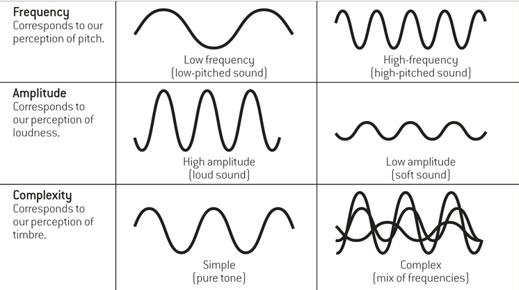
The frequency (or wavelength) of the sound wave depends on how often the peak in air pressure passes the ear or a microphone, measured in cycles per second, or hertz (Hz). Changes in the physical frequency of a sound wave are perceived by humans as changes in pitch, how high or low a sound is.
pitch
How high or low a sound is.
The amplitude of a sound wave refers to its height, relative to the threshold for human hearing (which is set at zero decibels, or dBs). Amplitude corresponds to loudness, or a sound’s intensity. The rustling of leaves in a soft breeze is about 20 dB, normal conversation is measured at about 40 dB, shouting produces 70 dB, a Slayer concert is about 130 dB, and the sound of the space shuttle taking off 1 mile away registers at 160 dB or more.
loudness
A sound’s intensity.
Differences in the complexity of sound waves, or their mix of frequencies, correspond to timbre, a listener’s experience of sound quality or resonance. Timbre (pronounced “TAM-
ber”) offers us information about the nature of sound. The same note played at the same loudness produces a perceptually different experience depending on whether it was played on a flute versus a trumpet, a phenomenon due entirely to timbre. timbre
A listener’s experience of sound quality or resonance.
Why does one note sound so different on a flute and a trumpet?
119
Most sounds—
The Human Ear
How does the auditory system convert sound waves into neural signals? The process is very different from the visual system, which is not surprising, given that light is a form of electromagnetic radiation, whereas sound is a physical change in air pressure over time: Different forms of energy require different processes of transduction. The human ear is divided into three distinct parts, as shown in FIGURE 4.21: the outer ear, the middle ear, and the inner ear.
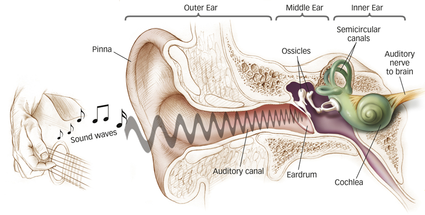
The outer ear consists of the visible part on the outside of the head (called the pinna); the auditory canal; and the eardrum, an airtight flap of skin that vibrates in response to sound waves gathered by the pinna and channeled through the canal. The middle ear, a tiny, air-
How do hair cells in the ear enable us to hear?
The inner ear contains the spiral-
cochlea
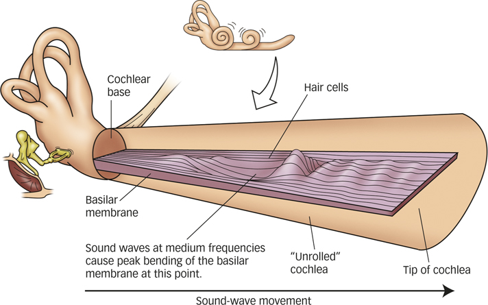
A fluid-
basilar membrane
A structure in the inner ear that undulates when vibrations from the ossicles reach the cochlear fluid.
hair cells
Specialized auditory receptor neurons embedded in the basilar membrane.
120
Perceiving Pitch
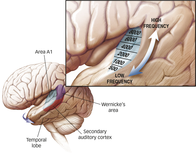
From the inner ear, action potentials in the auditory nerve travel to the thalamus and ultimately to an area of the cerebral cortex called area A1, a portion of the temporal lobe that contains the primary auditory cortex (see FIGURE 4.23). Neurons in area A1 respond well to simple tones, and successive auditory areas in the brain process sounds of increasing complexity (see FIGURE 4.23, inset; Rauschecker & Scott, 2009; Schreiner, Read, & Sutter, 2000; Schreiner & Winer, 2007). For most of us, the auditory areas in the left hemisphere analyze sounds related to language and those in the right hemisphere specialize in rhythmic sounds and music. There is also evidence that the auditory cortex is composed of two distinct streams, roughly analogous to the dorsal and ventral streams of the visual system. Spatial (“where”) auditory features, which allow you to locate the source of a sound in space, are handled by areas toward the back (caudal) part of the auditory cortex, whereas nonspatial (“what”) features, which allow you to identify the sound, are handled by areas in the lower (ventral) part of the auditory cortex (Recanzone & Sutter, 2008).
area A1
A portion of the temporal lobe that contains the primary auditory cortex.
How is the frequency of a sound wave encoded in a neural signal? Our ears have evolved two mechanisms to encode sound-
How does the frequency of a sound wave relate to what we hear?
The place code, used mainly for high frequencies, refers to the process by which different frequencies stimulate neural signals at specific places along the basilar membrane. Sounds of different frequencies cause waves that peak at different points on the basilar membrane (see FIGURE 4.22). When the frequency is low, the wide, floppy tip (apex) of the basilar membrane moves the most; when the frequency is high, the narrow, stiff end (base) of the membrane moves the most. The movement of the basilar membrane causes hair cells to bend, initiating a neural signal in the auditory nerve. Axons fire the strongest in the hair cells along the area of the basilar membrane that moves the most, and the brain uses information about which axons are the most active to help determine the pitch you “hear.”
place code
The process by which different frequencies stimulate neural signals at specific places along the basilar membrane, from which the brain determines pitch.
A complementary process handles lower frequencies. A temporal code registers relatively low frequencies (up to about 5000 Hz) via the firing rate of action potentials entering the auditory nerve. Action potentials from the hair cells are synchronized in time with the peaks of the incoming sound waves (Johnson, 1980). If you imagine the rhythmic boom-
boom- of a bass drum, you can probably also imagine the fire-boom fire- of action potentials corresponding to the beats. This process supplements the information provided by the place code.fire temporal code
The cochlea registers low frequencies via the firing rate of action potentials entering the auditory nerve.
121
Localizing Sound Sources
Just as the differing positions of our eyes give us stereoscopic vision, the placement of our ears on opposite sides of the head gives us stereophonic hearing. The sound arriving at the ear closer to the sound source is louder than the sound in the farther ear, mainly because the listener’s head partially blocks sound energy. This loudness difference decreases as the sound source moves from a position directly to one side (maximal difference) to straight ahead (no difference).
Another cue to a sound’s location arises from timing: Sound waves arrive a little sooner at the near ear than at the far ear. The timing difference can be as brief as a few microseconds, but together with the intensity difference, this time difference is sufficient to allow us to perceive the location of a sound. When the sound source is ambiguous, you may find yourself turning your head from side to side to localize the source. By doing this, you are changing the relative intensity and timing of sound waves arriving in your ears and collecting better information about the likely source of the sound. Turning your head also allows you to use your eyes to locate the source of the sound—
Hearing Loss
Broadly speaking, hearing loss has two main causes. Conductive hearing loss arises because the eardrum or ossicles are damaged to the point that they cannot conduct sound waves effectively to the cochlea. In many cases, medication or surgery can correct the problem. Sound amplification from a hearing aid also can improve hearing through conduction to the cochlea via the bones around the ear directly.
Sound amplification helps in the case of which type of hearing loss?
Sensorineural hearing loss is caused by damage to the cochlea, the hair cells, or the auditory nerve, and it happens to almost all of us as we age. Sensorineural hearing loss can be heightened in people regularly exposed to high noise levels (such as rock musicians or jet mechanics). Simply amplifying the sound does not help because the hair cells can no longer transduce sound waves. In these cases, a cochlear implant may offer some relief.
122
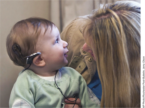
A cochlear implant is an electronic device that replaces the function of the hair cells (Waltzman, 2006). The external parts of the device include a microphone and speech processor, about the size of a USB key, worn behind the ear, and a small, flat, external transmitter that sits on the scalp behind the ear. The implanted parts include a receiver just inside the skull and a thin wire containing electrodes inserted into the cochlea to stimulate the auditory nerve. Sound picked up by the microphone is transformed into electric signals by the speech processor, which is essentially a small computer. The signal is transmitted to the implanted receiver, which activates the electrodes in the cochlea. Cochlear implants are now in routine use and can improve hearing to the point where speech can be understood.
Marked hearing loss is commonly experienced by people as they grow older, but is rare in an infant. However, infants who have not yet learned to speak are especially vulnerable because they may miss the critical period for language learning (see the Learning chapter). Without auditory feedback during this time, normal speech is nearly impossible to achieve, but early use of cochlear implants has been associated with improved speech and language skills for deaf children (Hay-
SUMMARY QUIZ [4.4]
Question 4.10
| 1. | What does the frequency of a sound wave determine? |
- pitch
- loudness
- sound quality
- timbre
a.
Question 4.11
| 2. | The placement of our ears on opposite sides of the head is crucial to our ability to |
- localize sound sources.
- determine pitch.
- judge intensity.
- recognize complexity.
a.
Question 4.12
| 3. | The place code works best for encoding |
- high intensities.
- low intensities.
- high frequencies.
- low frequencies.
c.
123
Hot Science: Music Training: Worth the Time
Music Training: Worth the Time
Did you learn to play an instrument when you were younger? If so, there’s good news for your brain. Compared to nonmusicians, musicians have greater plasticity in the motor cortex (Rosenkranz, Williamon, & Rothwell, 2007) and increased grey matter in motor and auditory brain regions (Gaser & Schlaug, 2003; Hannon & Trainor, 2007). But musical training also extends to auditory processing in nonmusical domains (Kraus & Chandrasekaran, 2010). Musicians, for example, show enhanced brain responses when listening to speech compared with nonmusicians (Parbery-
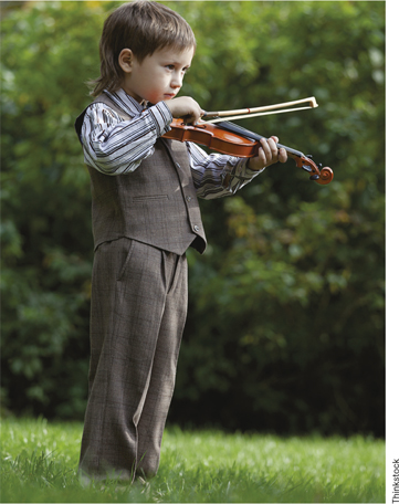
Remembering to be careful not to confuse correlation with causation, you may ask: Do differences between musicians and nonmusicians reflect the effects of musical training, or do they reflect individual differences, perhaps genetic ones, that lead some people to become musicians in the first place? Maybe people blessed with enhanced brain responses to musical or other auditory stimuli decide to become musicians because of their natural abilities. Recent experiments support a causal role for musical training. One study compared two groups of 8-
We don’t yet know all the reasons why musical training has such broad effects on auditory processing, but one likely contributor is that learning to play an instrument demands attention to precise details of sounds (Kraus & Chandrasekaran, 2010). Future studies will no doubt pinpoint additional factors, but the research to date leaves little room for doubt that your hours of practice were indeed worth the time.