2.6 The Amazing Brain
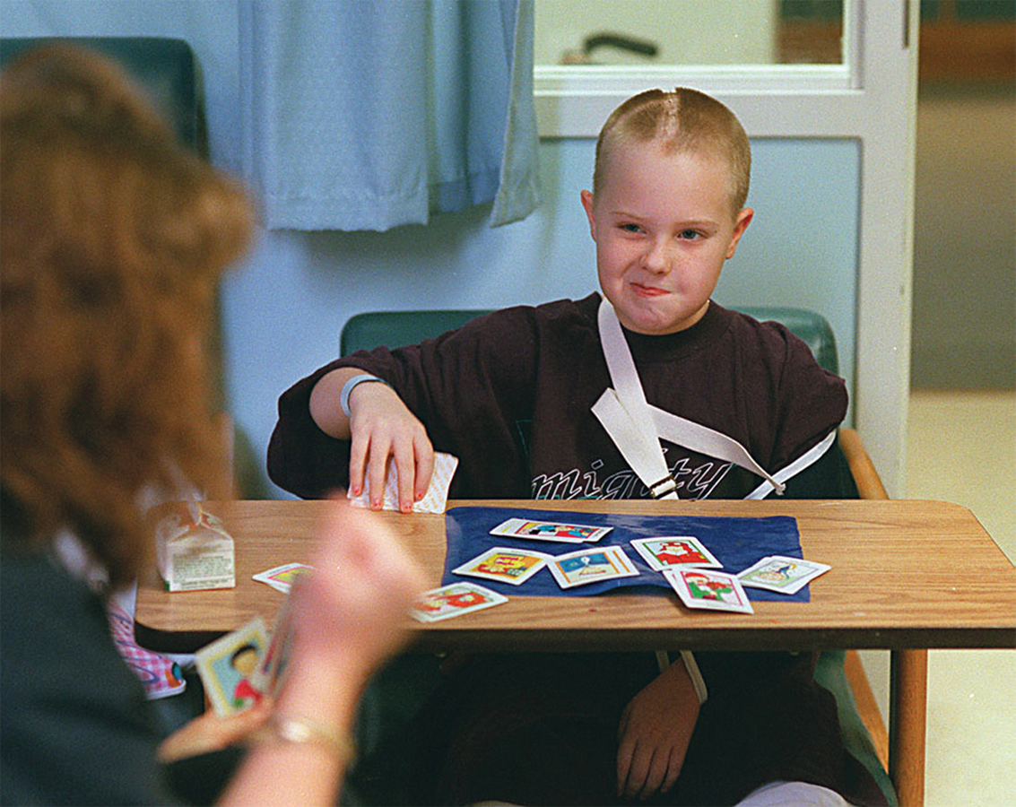
When Christina was wheeled out of surgery, her mother approached, grabbed hold of her right hand, and asked her to squeeze. Christina squeezed, demonstrating that she could understand and respond to language. Remember, she still had her left hemisphere.
Christina may not have lost her language abilities, but losing the right hemisphere did come at a cost. We know that Christina suffers partial paralysis on the left side of her body; this makes sense, because the right hemisphere controls movement and sensation on the left. We also know that it took Christina extra time to do her schoolwork. But if you ask Christina whether she has significant difficulty with any of the “right-brain” tasks described earlier, her answer will be no.
Christina’s recovery has been nothing short of astonishing. After the surgery, she went back to school and outperformed many of her classmates. In addition to making the honor roll and leading the bowling team, she managed to get her driver’s license (even though some of her doctors said she never would), graduate from high school, and go to college. These accomplishments are the result of Christina’s steadfast determination, but also a testament to the brain’s amazing ability to heal and regenerate.
Christina: What kind of therapy did you have and for how long?
Neuroplasticity
LO 11 Define neuroplasticity and recognize when it is evident in brains.
The brain undergoes constant alteration in response to experiences and is capable of some degree of physical adaptation and repair. Its ability to heal, grow new connections, and make do with what is available is a characteristic we refer to as neuroplasticity. New connections are constantly forming between neurons, and unused ones are fading away. Vast networks of neurons have the ability to reorganize in order to adapt to the environment and an organism’s ever-changing needs, a quality particularly evident in the young. After brain injuries, younger children have better outcomes than do adults; their brains show more plasticity (Johnston, 2009).
In one study, researchers removed the eyes of newborn opossums and found that brain tissues normally destined to become visual processing centers took a developmental turn. Instead, they became areas that specialize in processing other types of sensory stimuli, such as sounds and touch (Karlen, Kahn, & Krubitzer, 2006). The same appears to happen in humans. Brain scans reveal that when visually impaired individuals learn to read Braille early in life, a region of the brain that normally specializes in handling visual information becomes activated, suggesting it is used instead for processing touch sensations (Burton, 2003; Liu et al., 2007).
CONNECTIONS
In Chapter 1, we described the guidelines psychologists use to ensure the ethical treatment of humans and animals in their studies. In the study described here, the researchers were required to get approval from an ethics board to conduct their study. The board determined the proposed research required the surgery on the baby opossums and that they would be treated humanely.
Remarkably, this plasticity is evident even with the loss of an entire hemisphere. Researchers report that the younger the patient is when she has a hemispherectomy, the better her chances are for recovery. In fact, even with the loss of an entire left hemisphere (the primary location for language processing), speech is less severely impacted (though some impact is inevitable) in young patients. The younger the person undergoing the procedure, the less disability is evident in speech (Choi, 2008).
Stem Cells
Scientists once thought that people were born with all the neurons they would ever have. Brain cells might die, but no new ones would crop up to replace them. Thanks to research beginning in the 1990s, that dismal notion has been turned on its head. In the last few decades, studies with animals and humans have shown that some areas of the brain are constantly generating new neurons, a process known as neurogenesis, which might be tied to learning and creating new memories (Eriksson et al., 1998; Gould, Beylin, Tanapat, Reeves, & Shors, 1999; Reynolds & Weiss, 1992).

The cells responsible for churning out these new neurons are known as stem cells, and they are quite a hot topic in biomedical research. Scientists hope to harness these little cell factories to repair tissue that has been damaged or destroyed. Imagine that you could use stem cells to bring back all the neurons that Brandon lost from his injury or replace those that Christina lost to surgery. Cultivating new brain tissue is just one potential application of stem cell science. These cellular cure-alls might also be used to alleviate the symptoms of Parkinson’s disease, or replenish neurons of the spine, enabling people with spinal cord injuries to regain movement. Both have already been accomplished in mice (Keirstead et al., 2005; Wernig et al., 2008). Although embryonic stem cells can be used for such purposes, some stem cells can be found in tissues of the adult body, such as the brain and bone marrow.
Earlier we explained how neuroplasticity is more evident in young people. So what can we do to promote positive brain changes in our children? Some people think that early exposure to music is a critical factor, but is there evidence to support this belief?
across the WORLD
The Plastic Brains of Our Children
 Some 40 million children in China are learning to play the piano. Special piano kindergartens have sprung up across that country, their 4- and 5-year-old pupils studying scales and chords on top of traditional subjects. After school, these children may devote another 2 to 3 hours to piano practice (Lebrecht, 2010, February 1; Trelawny, 2008, June 5). The reason many Chinese parents push the piano so hard? Learning an instrument, they believe, will help their children thrive in school and life (Trelawny, 2008, June 5).
Meanwhile on the other side of the Pacific, American parents are playing classical music to their unborn fetuses and showing “Baby Mozart” DVDs to their babies and toddlers. Many of these moms and dads have been led to believe that early exposure to classical music promotes brain development, and they will do whatever it takes to give their kids a leg up on their peers (Hirsh-Pasek, Golinkoff, & Eyer, 2003).
DOES MUSICAL TRAINING MAKE YOU SMARTER?
Some evidence suggests that learning an instrument may indeed have cognitive benefits. Studies show that people with musical training generally score higher on language, auditory, and overall IQ tests than those without such training (Schellenberg, 2011; Schellenberg & Winner, 2011). But such research is correlational; the link between the variables is not necessarily causal. Even with a strong correlation or link between playing a musical instrument and increased cognitive abilities, we cannot be sure that one of these variables causes changes in the other. The only way to determine a cause-and-effect link is to conduct a study using the experimental method. As for playing Mozart to babies, there is no solid data demonstrating that merely listening to music enhances intelligence (Hirsh-Pasek et al., 2003). Enjoying the dynamic melodies of Mozart stimulates the brain and may improve mood, which can provide temporary cognitive benefits, but the same could be said for a host of other activities (Schellenberg, 2011).
CONNECTIONS
In Chapter 1, we discussed the problems with assuming a correlation between two variables can demonstrate a causal relationship. In this example, we cannot assume that music playing causes increased cognitive abilities. The direction may be the opposite (increased cognitive abilities leads to more interest in playing music) or some third variable may be involved (higher socioeconomic status linked to both music and better scores on tests).
From Bumps to Brain Scans: We’ve Come a Long Way
Both Brandon and Christina underwent many brain scans before and after their surgeries, which allowed doctors to get a detailed look inside their heads without lifting a scalpel. But had Brandon and Christina lived in a different era, brain scans would not have been an option.
CONNECTIONS
If phrenology were practiced today, we would consider it a pseudoscience, or an activity that resembles science but rests upon false assumptions and is unsubstantiated (see Chapter 1 to read more on pseudoscience).
Before there were technologies to study the brain, people could only speculate about what was going on beneath the skull of a living person. One theory was that bumps on a person’s skull could reveal characteristics about him. Judging the topography of a person’s head was a core part of phrenology, the now discredited brain “science” that achieved enormous popularity at the beginning of the 19th century through its founder Franz Joseph Gall (1757–1828). But flaws in the theory behind phrenology were recognized early on. Another early (but more scientific) way of studying the brain was through ablation, a technique used by physiologist Pierre Flourens (1794–1867) to determine the functions of different brain regions (Pearce, 2009). This technique involved destroying parts of the brain of living animals and then determining whether some functioning was lost following this surgery.
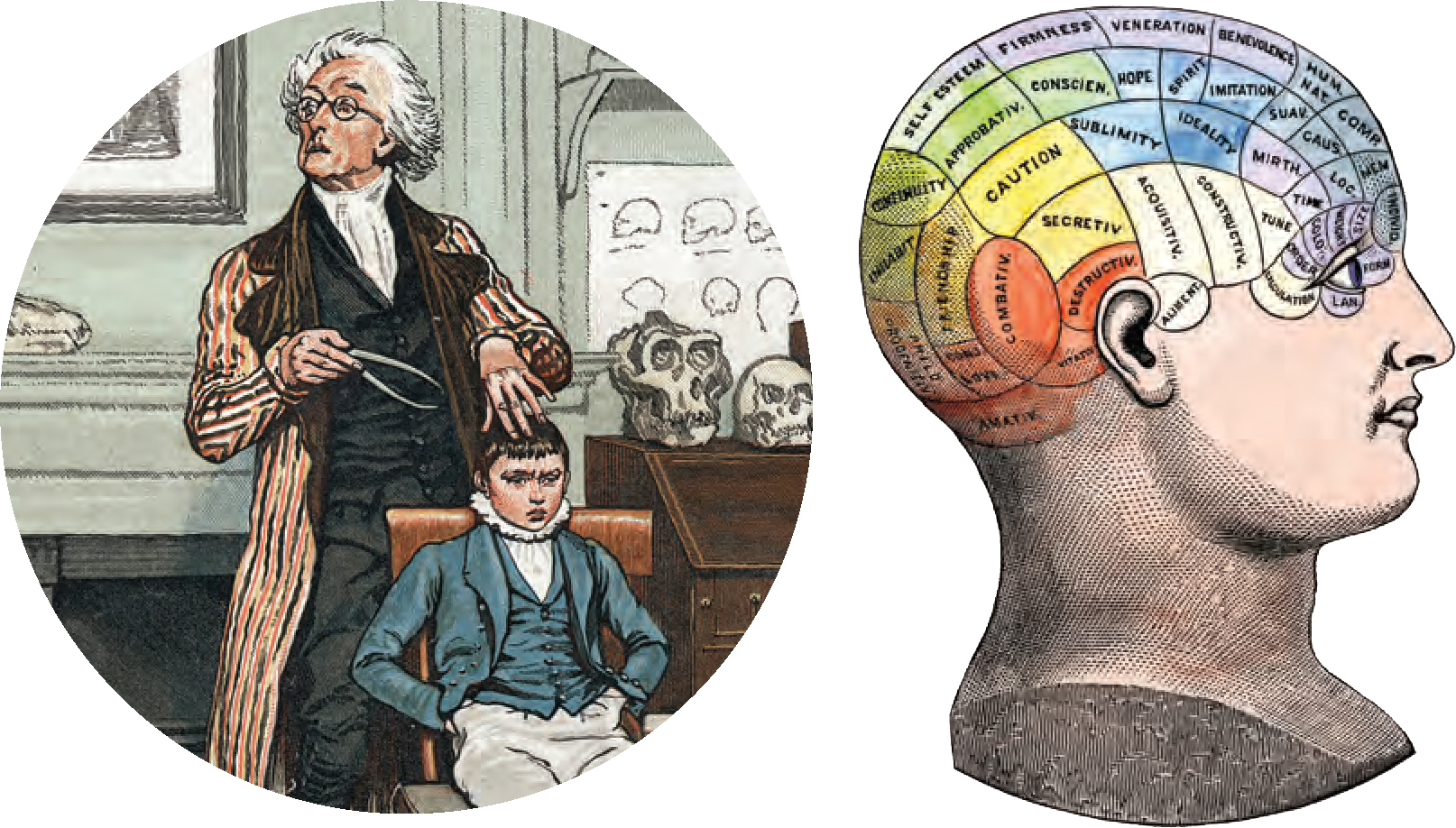
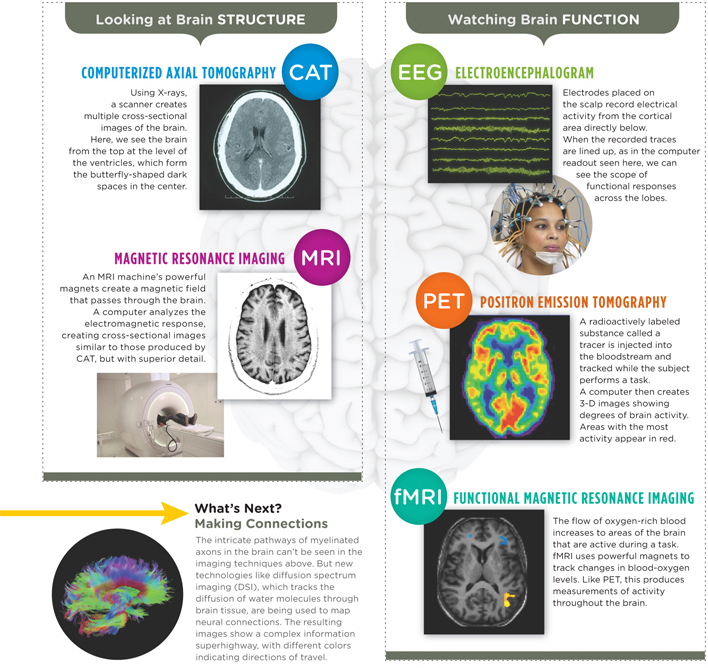
These days, scientists no longer try to interpret what’s going on inside living brains by running fingers over people’s heads. There are now technologies that allow us to sift through the brain’s constant buzz of activity, behold its three-dimensional structure, and identify interesting hot spots (TABLE 2.1). Nevertheless, one of Gall and Flourens’ major contributions was the idea that there might be areas of the brain that have particular functions. In other words, there are locations in the brain that are responsible for specific brain activities: there is a localization of function.
| Function | Use | Limitations | |
| Electroencephalogram (EEG) | Detects electrical energy in the brain and displays information that can be interpreted. | Monitors and studies brain processes, diagnoses brain conditions, and sheds light on the way substances affect brain function. | Records activity happening on the surface of the brain. |
| Computerized Axial Tomography (CAT scan or CT scan) | X-rays create many cross-sectional images of the brain (or body); similar to bread slices, you can examine the pieces individually. | Spots tumors and brain damage and presents the brain’s structural features. | Exposes people to radiation, potentially increasing cancer risk (National Cancer Institute, 2010). |
| Positron Emission Tomography (PET) | Uses radioactive glucose; scanner sensitive to radioactivity, detects active areas of the brain, which have absorbed the most sugar. | Captures the brain in a variety of activities (e.g., dreaming, hallucinating, or appreciating the scent of a rose). | From injection to scan, PET scans are time-consuming. This procedure can be expensive, and it exposes people to radiation. |
| Magnetic Resonance Imaging (MRI) | Using radio frequency waves, the MRI produces cross-sectional images (slices) of the brain; detects tumors, bleeding, infections, and the size and shape of various brain structures. | Provides information on the anatomy of the brain. | MRIs produce more detailed images than CTs, but are more time-consuming and expensive. |
| Functional Magnetic Resonance Imaging (fMRI) | Captures changes in brain activity. | Reveals patterns of blood flow in a particular area of the brain, providing a good indicator of how much oxygen is being used as a result of activity. | Indirectly measures neural activity via blood flow, not necessarily identifying the precise location of cognitive processes. |
| Technologies to study the brain are continuing to evolve. Summarized here are the most commonly used technologies. | |||
| SOURCE: YASMIN ET AL., 2012. | |||
SOCIAL MEDIA and psychology
 One of the main themes of this chapter is specialization, the idea that certain areas of the brain tend to become active during certain tasks. When we say “tasks,” we mean just about any activity you can imagine, from riding a bicycle to managing friend networks on Facebook.
The average number of Facebook “friends” is 245, but the friend tally varies significantly from one person to the next, ranging from zero to 5,000, the maximum allowed by Facebook (Ball, 2010, May 28; Tsukayama, 2012, February 3). What does a Facebook friend number reveal about a person—job networking skills, offline popularity, time wasted at work? According to one preliminary study, friend volume may reflect something about the brain of the user.
Using MRI technology, researchers studied the brain structures of a sample of Facebook users. What they discovered was a correlation between the number of Facebook friends and the density of gray matter (the primary tissue of the cerebral cortex) in areas of the brain important for social interaction. One of those regions, the superior temporal sulcus, is thought to be important for detecting socially meaningful movements such as hand gestures and eye shifts. Another, known as the entorhinal cortex, appears to play a key role in matching faces to names, a critical skill for Facebookers (Kanai, Bahrami, Roylance, & Rees, 2012). As anyone with a few hundred friends can testify, keeping track of all those names and faces can be challenging.
FACEBOOK FRIENDS: A GRAY MATTER.
We should point out that this study is merely correlational. It does not reveal whether the number of friends causes changes in brain structure, or whether the characteristics of brain structures determine the number of friends. Perhaps some other variable is responsible for both. This single study needs replication, but it has generated intriguing questions for researchers to tackle in the future.
LO 12 Compare and contrast tools scientists use to study the brain.
In the past, most of what scientists learned about the brain came from probing the skulls of cadavers and observing the behavior of people suffering from brain damage. But the last century, and particularly the last few decades, has witnessed an explosion of brain research technologies. Such technological advances have made it possible to observe the brain as it makes decisions, sleeps, and even tells lies (Dang-Vu et al., 2008; Hampton & O’Doherty, 2007; Kozel et al., 2009). See TABLE 2.1 for information on the different types of technologies, their function, uses, and limitations (also see Infographic 2.3). With the technology available today, we can even track the activities of strings of neurons.
from the pages of SCIENTIFIC AMERICAN
Electric Surprise
Stimulating brain cells may be trickier than we thought
Scientists and doctors have long used electricity to both study and treat the brain. But a report in the August 27, 2009, issue of Neuron indicates that the brain’s response to electricity is exceptionally complex. Using a new type of optical imaging, Harvard Medical School researchers observed neurons as they were stimulated by an electrode. Instead of activating a small sphere of surrounding neurons as expected, the electrodes caused sparse strings of neurons to fire across the brain. The finding suggests that brain surgeons and the designers of neural prosthetics have a much smaller margin of error than previously thought—shifting an electrode even slightly could activate an entirely different set of neurons. Melinda Wenner. Reproduced with permission. Copyright © 2010 Scientific American, a division of Nature America, Inc. All rights reserved.
INFOGRAPHIC 2.3: Ways to Study the Living Brain
For hundreds of years, scientists interested in the brain were limited to surgical techniques, often on cadavers. Imaging and recording technologies now allow us to investigate the living brain by assessing structure, function, or both. CAT and MRI techniques provide static pictures of brain structures, while functional imaging and recording techniques allow us to see the relationship between brain activity and specific mental functions. Functional techniques can also be used to diagnose injuries and diseases earlier than techniques that look at structure.
New technologies are continually being developed, allowing us to study the brain in ways we couldn’t imagine just a few years ago.
show what you know
Question 2.16
1. Scientists hope that in the future they will be able to discover how we can use stem cells to help people like Brandon and Christina. The goal would be for doctors to induce the process of __________ to restore the lost brain tissue.
- ablation
- agonists
- neurogenesis
- lateralization
c. neurogenesis
Question 2.17
2. You have been asked to set up an experiment to determine if playing classical music to infants leads to improved cognitive abilities. What would your independent and dependent variables be? How would your experimental and control groups be treated differently?
Independent variable: manipulation of hearing classical music.
Dependent variable: measure of cognitive abilities.
Answers will vary. The experimental group would listen to classical music and the control group would not listen to classical music.
Question 2.18
3. __________ are responsible for creating new neurons.
Stem cells
Question 2.19
4. The brain is constantly undergoing alterations in response to experiences and is capable of a certain degree of physical adaptation and repair. This ability is known as:
- neuroplasticity
- phrenology
- ablation
- lateralization
a. neuroplasticity