2.7 The Cortex: A Peek Beneath the Skull
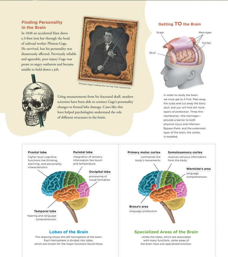
Imagine you were one of the surgeons performing Christina’s hemispherectomy. What exactly would you see when you peeled away the scalp and cut an opening into the skull? Before seeing the brain, you would come upon a layer of three thin membranes, the meninges, which envelop and protect the brain and spinal cord. Perhaps you have heard of meningitis, a potentially life-threatening condition in which the meninges become inflamed as a result of an infection. The meninges are bathed in a clear watery substance called cerebrospinal fluid, which offers additional cushioning and helps in the transport of nutrients and waste in and out of the brain and the spinal cord. Once you peeled back the meninges, you would behold the pink cerebrum.
Cerebral Cortex
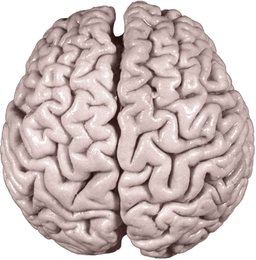
As Christina’s surgeon, your main task would be to remove part of the cerebrum’s outermost layer, the cerebral cortex (sə-rē-brəl). The cerebral cortex processes information and is the layer of cells surrounding nearly all the brain’s structures. You’ll remember from our earlier presentation that the cerebrum looks like a wrinkled walnut. This is because the cortex is scrunched up and folded onto itself to fit inside a small space (the skull).
The Lobes of the Brain
LO 13 Identify the lobes of the cortex and explain their functions.
The cortex overlaying each hemisphere is separated into different sections, or lobes (Infographic 2.4). The major function of the frontal lobes is to organize information among the other lobes of the brain. The frontal lobes are also responsible for higher-level cognitive functions, such as thinking, perception, and impulse control. The parietal lobes (pə-rī-ə-təl) receive and process sensory information such as touch, pressure, temperature, and spatial orientation. Visual information goes to the occipital lobes (äk-si-pə-təl) for processing, and hearing and language comprehension are largely handled by the temporal lobes. We’ll have more to say about the lobes shortly.
The Association Areas
LO 14 Recognize the association areas and identify their functions.
In addition to these broad functions of the brain lobes, there are specific functions associated with particular areas of the lobes. Some areas in the lobes are home to networks of neurons that specialize in certain activities. The motor areas direct movement, the sensory areas receive and analyze sensory stimuli, and the association areas integrate information from all over the brain. The association areas are located in all four lobes of the brain; however, they are much harder to pinpoint than are the motor and sensory areas. The association areas allow us to learn (just as you’re doing now), have abstract thoughts (for example, 2 + 2 = 4), and carry out complex behaviors like texting and tweeting. The language-processing hubs we learned about earlier, Broca’s area and Wernicke’s area, are association areas that play a role in the production and comprehension of speech. In humans, the vast majority of the cortical surface is dedicated to the association areas.
The Lobes: Up Close and Personal
Prior to her hemispherectomy, Christina was extroverted, easygoing, and full of energy. “I had absolutely no worries,” she says, recalling her pre-Rasmussen’s days. After her operation, Christina became more introverted and passive. She felt more emotionally unsettled. “You go into surgery one person,” she says, “and you come out another.”
The transformation of Christina’s personality may be a result of many factors, including the stress of dealing with a serious disease, undergoing a major surgery, and readjusting to life with disabilities. But it could also have something to do with the fact that she lost a considerable amount of brain tissue, including her right frontal lobe. Networks of neurons in the frontal lobes are involved in processing emotions, making plans, controlling impulses, and carrying out a vast array of mental tasks that each person does in a unique way (Williams, Suchy, & Kraybill, 2010). The frontal lobes play a key role in the development of personality (Stuss & Alexander, 2000). A striking illustration of this phenomenon involves an unlucky railroad foreman, Phineas Gage.
Phineas Gage and The Frontal Lobes
The year was 1848, and Phineas Gage was working on the railroad. An accidental explosion sent a 3-foot iron tamping rod clear through his frontal lobes (Infographic 2.4). The rod, about as thick as a broom handle, drove straight into Gage’s left cheek, through his brain, and out the top of his skull (Macmillan, 2000). What’s peculiar about Gage’s mishap (besides the fact that he was walking and talking just hours later) is the extreme transformation it caused. Before the accident, Gage was a well-balanced, diligent worker whom his supervisors referred to as their “most efficient and capable foreman” (Harlow, 1848, as cited in Neylan, 1999, p. 280). After the accident, he was unreliable, unpleasant, and downright vulgar. His character was so altered that people acquainted with him before and after the accident claimed he was “no longer Gage” (Harlow, 1848, as cited in Neylan, 1999, p. 280). As he could not continue working as a foreman, Gage eventually began to travel to the larger towns of New England, “exhibiting himself and the tamping iron” (Macmillan, 2000, p. 55).
Modern scientists have revisited Gage’s case, using measurements from his fractured skull and brain imaging data to estimate exactly where the damage occurred. Their studies suggest that the metal rod caused destruction in both the left and right frontal lobes (Damasio, Grabowski, Frank, Galaburda, & Damasio, 1994). The only good thing about Gage’s horrible accident, it seems, is that it illuminated the importance of the frontal lobes in defining personality characteristics.
Canines and The Motor Cortex
Toward the rear of the frontal lobes is a strip of the brain known as the motor cortex, which works with other areas to plan and execute voluntary movements (Infographic 2.4). Persuasive evidence pointing to this region’s involvement in muscle movement came initially from a study of dogs by Gustav Fritsch (1838–1927) and Edvard Hitzig (1838–1907). Working from a makeshift lab, the two doctors discovered they could make the animals move by electrically stimulating their brains (Gross, 2007). A mild shock to the right side of the cortex might cause a twitch in the left forepaw or the left side of the face, whereas stimulating the left would spur movement on the right (Finger, 2001).
Albert Einstein and The Parietal Lobes
CONNECTIONS
This research study compared one man’s brain to a control group of 35 men. In Chapter 1, we discussed the importance of having a large enough sample size to give us reliable findings, which might be a consideration here. Also, we discussed the potential problems with using case studies for making generalizations to the population, and in the current study, the use of Einstein as a single participant to compare to the control group could be considered a type of case study.
Directly behind the frontal lobes on the crown of your head are the parietal lobes (Infographic 2.4). The parietal lobes help orient the body in space, are involved in tactile processing (for example, interpreting sensations related to touch, such as pain and pressure), and may play a role in mathematical reasoning associated with spatial cognition (Grabner et al., 2007; Wolpert, Goodbody, & Husain, 1998). Let’s take a closer look at one study comparing the brain of Albert Einstein to a control group of 35 brain specimens from men who had donated their bodies for use in research. Prior to their deaths, these men had normal cognitive functioning, average intelligence, and no mental health issues. The researchers reported that Einstein’s brain did not weigh more than the average brain of the control group, but a region of his parietal lobe believed to be important for visual–spatial processing was 15% larger than those of the control group. They proposed that the differences in that specific region of the parietal lobe in Einstein’s brain may be linked to his “exceptional intellect” in areas of visuospatial cognition and mathematical thinking (Witelson, Kigar, & Harvey, 1999). But again, the link between the size of the parietal region and “exceptional intellect” is correlational in nature, so we should be cautious in our interpretations.
INFOGRAPHIC 2.4: Getting Into the Brain
Penfield, The Homunculus, and The Somatosensory Cortex
North American neurosurgeon Wilder Penfield (1891–1976) used a method similar to Fritsch and Hitzig with humans to create a map showing which points along the motor cortex corresponded to the various parts of the body (Penfield & Boldrey, 1937). Penfield’s map is often represented by the oversimplified “homunculus” (hō′məη-kyə-ləs; Latin for “little man”) cartoon, a distorted image of a human body with huge lips and hands and a tiny torso (Figure 2.9). The size of each body part roughly reflects the amount of cortex devoted to it, which explains why parts requiring extremely fine-tuned motor control (the lips and hands) are gigantic in comparison to the other body parts in the figure.
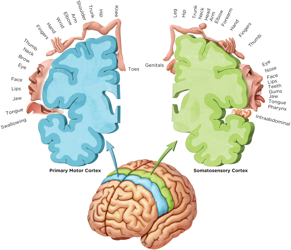
The parietal lobes are home to the somatosensory cortex, a strip of the brain running parallel to the motor cortex that receives and integrates sensory information from all over the body, for example, pain and temperature. Penfield, the neurosurgeon who created the homunculus for the primary motor cortex, mapped the somatosensory cortex in the same way (Penfield & Boldrey, 1937; Figure 2.9). As you might expect, the most sensitive areas of the body like the face and tongue are oversized on the homunculus, whereas areas less sensitive to stimulation, such as the forearm and the calf, are smaller.
The Temporal Lobes and The Auditory Cortex
Behind the parietal lobes, on the sides of your head, are the temporal lobes, which process auditory stimuli, recognize visual objects, especially faces, and play a key role in language comprehension and memory (Hickok & Poeppel, 2000; Infographic 2.4). The temporal lobes are home to the auditory cortex, which receives information from the ears and allows us to “hear” sounds. In particular, researchers have supported the notion, based on studies of primate vocalizations, that the ability to recognize language has evolved over time and is processed within the temporal lobes (Scott, Blank, Rosen, & Wise, 2000; Squire, Stark, & Clark, 2004).
The Occipital Lobes and The Primary Visual Cortex
Visual information is initially processed in the occipital lobes, in the lower back of the head (Infographic 2.4). If you have ever suffered a severe blow to the rear of your head, you may remember “seeing stars,” probably because activity in the occipital lobes was disrupted (hopefully only for a few seconds). It is here, where the optic nerve connects to the primary visual cortex, that our visual information is received and interpreted, and where we process information (for example, about color, shape, and motion). (See Chapter 3 on sensation and perception for a full discussion.) Even for a person with healthy eyes, if this area were damaged, severe visual impairment could occur. At the same time, the individual would still be able to “see” vivid mental images (Bridge, Harrold, Holmes, Stokes, & Kennard, 2012).
The cortex is the outermost section of the brain. It is also the part that is “newest,” or most recently evolved compared to the “older” structures closer to the core. We know this because researchers have compared the brains of humans with other primates. Those structures we share with our primate relatives are considered more primitive, or less evolved.
Now that we have surveyed the brain’s outer terrain, identifying some of the hotspots for language and other higher cognitive functions, let’s dig deeper and examine some of its older structures.
show what you know
Question 2.20
1. The major function of the __________ is to organize information among the other lobes of the brain.
- parietal lobes
- frontal lobes
- corpus callosum
- temporal lobes
b. frontal lobes
Question 2.21
2. The __________ integrate information from all over the brain, allowing us to learn, have abstract thoughts, and carry out complex behaviors.
association areas
Question 2.22
3. This section introduces the lobes of the cortex and their associated functions. Create a way to remember the lobes and their specific functions using a rhyme or another memorization technique you know.
Answers will vary. There is no right or wrong mnemonic; use one that will help you remember.
Here is one example connecting letters or word segments to function:
Frontal lobes: Front and center (integrating information from all over the brain)
Occipital lobes: “Oh, did you see that?” (visual processing)
Parietal lobes: Perceive touch (processing of touch sensations)
Temporal lobes: Tempo is just right (auditory processing)
Question 2.23
4. Phineas Gage suffered a horrific accident in the mid-1800s, when an explosion sent a metal rod through his brain and out the top of his skull. Which of the following caused the sudden change in his personality following the accident?
- damage to his occipital lobes
- damage to his temporal lobes
- damage to his frontal lobes
- damage to his somatosensory cortex
c. damage to his frontal lobes