3.2 Vision

Emma, Zoe, and Sophie were born over 3 months early, long before fetal development had run its course. Premature infants are at risk for serious complications, including respiratory distress, heart irregularities, and vision problems (March of Dimes, 2013).
 THE EYES OF THE TRIPLETS When Liz became pregnant with triplets, she was busy caring for her firstborn child, 3-
Many triplets are fraternal, meaning they come from three distinct egg–
After spending their first months of life in the hospital, hooked to feeding tubes, breathing tubes, and IVs, the triplets finally came home—
What happened to the triplets’ eyes? To understand how the girls lost their vision, we must first learn how the eye turns light into electrical and chemical impulses for the brain to interpret. 
Light Is Sight
LO 4 Explain how electromagnetic energy is transduced into a sensation of vision.
When you look at a stop sign, would you believe you are sensing light waves bouncing off the stop sign and into your eyes? The eyes do not sense faces, objects, or scenery. They detect light. Remember, if you don’t have light, you don’t have sight. But what exactly is light? Light is an electromagnetic energy wave, composed of fluctuating electric and magnetic fields zooming from place to place at a very fast rate. And when we say “fast,” we mean from Atlanta to Los Angeles in a tenth of a second. Electromagnetic energy waves are everywhere all the time, zipping past your head, and bouncing off your nose. As you can see in Figure 3.1, light that is visible to humans falls along a spectrum, or range, of electromagnetic energy.
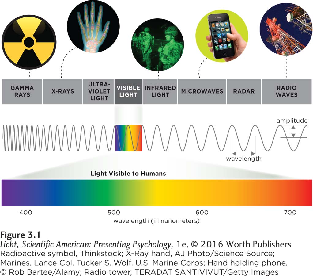
Only a small part of the electromagnetic spectrum can be detected by the human visual system. Visible light wavelengths range from approximately 400 to 700 nanometers. We use electromagnetic energy for a variety of purposes, from warming our dinners to carrying on digital conversations.
wavelength The distance between wave peaks (or troughs).
WAVELENGTH The various types of electromagnetic energy can be distinguished by their wavelength, which is the distance from one wave hump to the next (like the distance between the crests of waves rolling in the ocean; Figure 3.1). Gamma waves have short wavelengths and are located on the far left of the spectrum. At the opposite extreme (far right of the spectrum) are the long radio waves. The light humans can see falls in the middle of the spectrum, measuring between 400 and 700 nanometers (nm) or billionths of a meter (Brown & Wald, 1964). Wavelength also plays an important role in determining the colors humans and animals can detect.
The Colors We See

This is what you might perceive if you were a snake searching for dinner in the dark. The western diamondback rattlesnake has facial sensors that detect infrared radiation from warm-
Although dogs can only see the world in blues, yellows, and grays (Coren, 2008, October 20), primates, including humans, can detect a wider spectrum of colors, including reds and oranges. This ability to see reds and oranges may be an adaptation to spot ripe fruits against the green backdrop of tree leaves (Rowe, 2002). Other creatures can see “colors” that we can’t. Snakes can detect infrared waves radiating off the bodies of their prey, and birds size up potential mates using the ultraviolet waves reflected by feathers (Bennett, Cuthill, Partridge, & Maier, 1996; Gracheva et al., 2010).
hue The color of an object, determined by the wavelength of light it reflects.
amplitude The height of a wave; the distance from midpoint to peak, or from midpoint to the trough of a wave.
saturation Color purity.
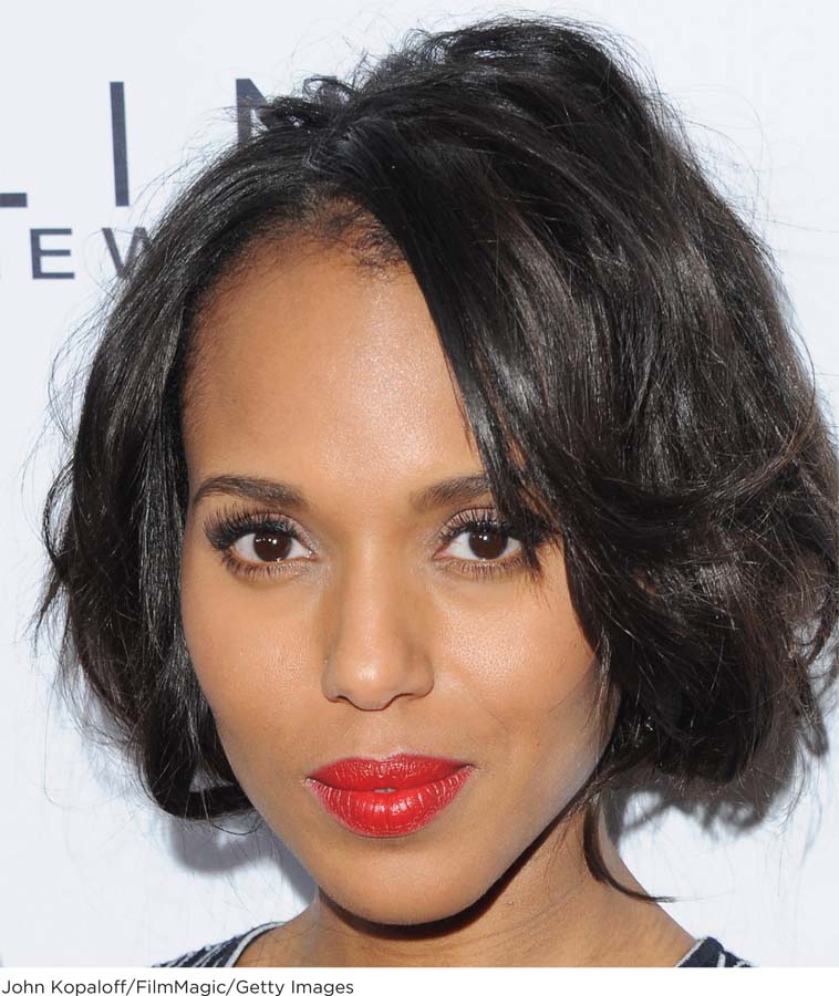
Kerry Washington sported intensely red lipstick at the Daily Front Row’s 2015 Annual Fashion Los Angeles Awards. Her lipstick may appear to be a pure red, but the color is most likely a blend of various red wavelengths. Most colors we encounter in the real world are a mix of different wavelengths.
FEATURES OF COLOR The colors you see result from light reflecting off of objects and reaching your eyes. Every color can be described according to three factors: hue, brightness, and saturation. The first factor, hue, is what we commonly refer to as “color” (blue jeans have a blue hue). Hue is determined by the wavelength reflecting off of an object: Violet has the shortest wavelength in the visible spectrum (400 nm), and red has the longest (700 nm). The brightness of a color represents a continuum from bright to dim, or the intensity of the color. Brightness depends on wave height, or amplitude, the distance from midpoint to peak (or from midpoint to trough; Figure 3.1). Just remember, the taller the height, the brighter the light. Saturation, or color purity, is determined by uniformity of wavelength. Saturated colors are made up of same-
PERCEPTION OF COLORS And now, the 2.3 million-
Go find the brightest, most saturated object you possess that has a yellow hue, and grab a strong flashlight while you are at it. Wait until it is dark outside. Now put the object on a table right in front of you and look at it with a dim light shining overhead. Next, shine the flashlight directly onto the yellow object and notice how your perception of the color changes. Finally, turn off all the lights in the room and notice again how your perception of the color changes.
try this
Your three perceptions of this same yellow object will be very different—
You Won’t Believe Your Eyes
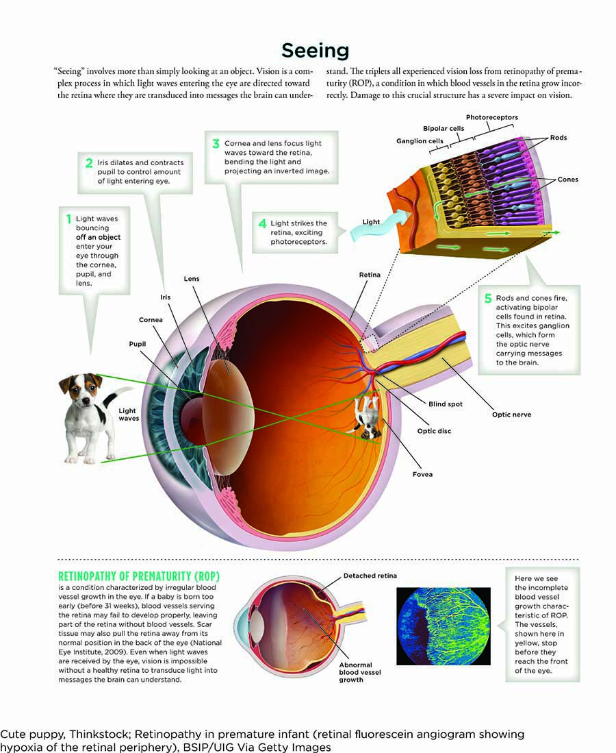
The human eye is nothing short of an engineering marvel (Infographic 3.2). When you look in the mirror, you see only a fraction of each ping-
cornea The clear, outer layer of the eye that shields it from damage and focuses incoming light waves.
THE CORNEA The surface of the eye looks wet and glassy. This clear outer layer over the colored portion of the eye is called the cornea, and it has two important jobs: (1) shielding the eye from damage by dust, bacteria, or even a poke, and (2) focusing incoming light waves. About 65–
iris The muscle responsible for changing the size of the pupil.
THE IRIS AND THE PUPIL Directly behind the cornea is the donut-
accommodation The process by which the lens changes shape in order to focus on images near and far.
THE LENS AND ACCOMMODATION Behind the pupil is the lens, a tough, transparent structure that is similar in size and shape to an “M&M’s candy” (Mayo Clinic, 2014). Like the cornea, the lens specializes in focusing incoming light, but it can also change shape in order to adjust to images near and far, a process called accommodation. If you take your eyes off this page and look across the room, faraway objects immediately come into focus because your lens changes shape. As we age, the lens begins to stiffen, impairing our ability to focus on up-
INFOGRAPHIC 3.2
The Retina
retina The layer of the eye that contains photoreceptor cells and the location for the transduction of light energy into neural activity.
After passing through the cornea, pupil, and lens, light waves travel through the eyeball’s jellylike filling and land on the retina, a carpet of neurons covering the back wall of the eye. This area was the source of the triplets’ visual impairments. Emma, Zoe, and Sophie all suffered from retinopathy of prematurity (ROP), a condition in which blood vessels in the retina grow incorrectly (Chawla et al., 2012). The last 3 months of pregnancy are critical for fetal eye development, when webs of blood vessels rapidly branch from the center of the retina outward, delivering crucial oxygen and nutrients to the developing tissue. After the triplets’ very early birth, these vessels began to branch abnormally, eventually pulling the retina from the back of the eye.
The retina is responsible for the transduction of light energy into neural activity; that is, sensing light and relaying a message to the brain. Without the retina, vision is impossible.
LO 5 Describe the function of rods and cones.
CONNECTIONS
In Chapter 2, we noted that neurons are activated in response to sensations. In the case of vision, the sensation is light and the neurons being stimulated start with the photoreceptors in the retina. When a photoreceptor fires, its message is sent on its way to the brain.
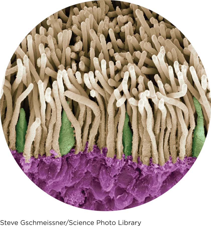
photoreceptors Cells that absorb light energy and turn it into chemical and electrical signals for the brain to process.
rods Specialized light receptors in the retina that are responsible for sight when the light level is low; not sensitive to color, but useful for night vision.
cones Specialized light receptors responsible for our sensation of color and our ability to sense details.
You can see why the light-
PHOTORECEPTORS AND OTHER NEURONS The retina is home to millions of specialized neurons called photoreceptors, which absorb light energy and turn it into chemical and electrical signals for the brain to process. Two types of photoreceptors are located along the back of the retina, rods and cones, which get their names from their characteristic shapes. Rods are extremely sensitive, firing in response to even a single photon, the smallest possible packet of light (Rieke & Baylor, 1998). If rods were all we had, the world would look something like an old black-
optic nerve The bundle of axons from ganglion cells leading to the visual cortex.
blind spot The location where the optic nerve exits the retina.
The rods and cones are just the first step in a complex neural signaling cascade that ultimately leads to a visual experience in the brain (see Infographic 3.2 on page 97). Near the rods and cones are bipolar cells, another specialized type of neuron, located approximately in the middle of the retina. When a rod or cone is stimulated by light energy, it conveys its signal to nearby bipolar cells. These in turn convey their signal to ganglion cells, yet another type of neuron, located toward the front of the retina. Axons of the ganglion cells bundle together in the optic nerve, which is like an electrical cable (one extending from each eye) hooking the retina to the brain. The optic nerve exits the retina at the optic disc, causing a blind spot, since this area lacks rods and cones. You can find your blind spot by following the instructions in the Try This.
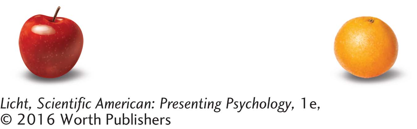
try this
Holding your book at arm’s length, close
your right eye and stare at the orange with your left
eye. Slowly bring the book closer to your face.
The apple on the left will disappear when light from
that picture falls on your blind spot.
THE FOVEA The retina in each eye is home to some 120 million rods and 6 million cones (Amesbury & Schallhorn, 2003). Rods are found everywhere in the retina, except in the optic disc (mentioned earlier) and a tiny central spot called the fovea. Cones are packed most densely in the fovea, but are also sprinkled through the rest of the retina. When you need to study something in precise detail (like the tiny serial number on the back of a computer), hold it under a bright light and stare at it straight-
Dark and Light Adaptation
dark adaptation Ability of the eyes to adjust to dark after exposure to brightness.
The eye has an amazing ability to adjust to drastic fluctuations in light levels. This process starts with the pupil, which rapidly shrinks and expands in response to light changes, and then continues with the rods and cones, which need more time to adjust to changes in lighting. When you walk into a dark movie theater after being outside in the bright sun, you can barely see an inch in front of your face. After a few minutes, your eyes start to adjust to the dark in a process called dark adaptation, which takes about 8 minutes for cones and 30 minutes for rods (Hecht & Mandelbaum, 1938; Klaver, Wolfs, Vingerling, Hoffman, & de Jong, 1998). Cones respond more quickly, so they are more useful in the first few minutes of dark adaptation. Then the rods kick into action, allowing you to make out silhouettes of objects and people. Why are these photoreceptors so sluggish in their responses to darkness? To restore their sensitivity to light, rods and cones must undergo a chemical change associated with protein molecules, and this takes time (Caruso, 2007, August 13; Ludel, 1978). Also keep in mind that, for most of human evolution, dark adaptation meant adjusting to the gradual setting of the sun (Caruso, 2007, August 13).
light adaptation Ability of the eyes to adjust to light after being in the dark.
When you leave the dark theater and return to the blinding light of day, the eyes also adjust. With light adaptation, the pupil constricts to reduce the amount of light flooding the retina, and the rods and cones become less sensitive to light. Light adaptation occurs relatively quickly, lasting at most 10 minutes (Ludel, 1978).
Is That Oprah Winfrey Over There?
Let’s stop for a moment and examine how information flows through the visual pathway (Infographic 3.2 on page 97). Suppose you are looking at Oprah Winfrey’s face. Remember, you’re actually seeing the light that her face reflects. Normally, light rays bouncing off Oprah would continue moving along a straight-
CONNECTIONS
Interneurons are found in the central nervous system (the brain and the spinal cord). As described in Chapter 2, the interneurons receive and help process signals from sensory neurons.
feature detectors Neurons in the visual cortex specialized in detecting specific features of the visual experience, such as angles, lines, and movements.
The optic nerves (one from each eye) intersect at a place in the brain called the optic chiasm. From there, information coming from each eye gets split, with about half traveling to the same-
In Living Color
Although we have discussed various features of color, we have not yet explained how waves of electromagnetic energy result in our perception of colors. How does the brain know red from maroon, green from turquoise, yellow from amber? Two main theories explain human color vision—
LO 6 Compare and contrast the theories of color vision.
trichromatic theory (trī-krō-ˈma-
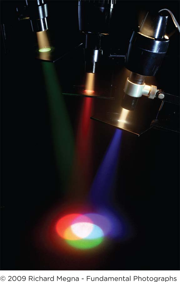
The color of a light is the result of a mixture of wavelengths from the visible spectrum. When red, blue, and green light wavelengths are combined in equal proportions, they produce white light. This may seem counterintuitive, because most of us have learned that mixing different-
THE TRICHROMATIC THEORY Proposed in the 1800s by an English physician–
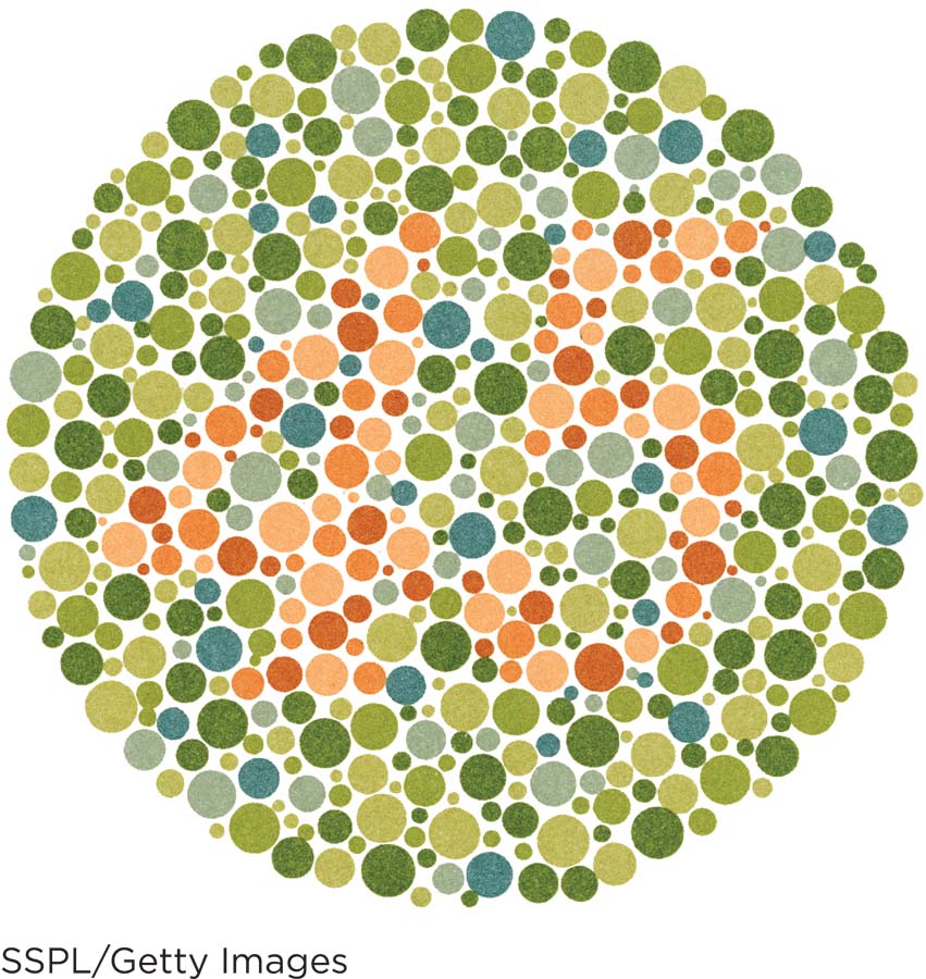
If you cannot make out the number 45 in this Ishihara color plate, then you might have a red–
So how is it that we can detect millions of colors when our cones are only sensitive to red, green, and blue? The primary colors of light are also red, green, and blue, and when mixed together in equal proportions, they appear as white light (see the photograph to the left). According to the trichromatic theory, the brain identifies a precise hue by calculating patterns of excitement among the three cone populations. When you look at a yellow banana, for example, both the red and green cones fire, but not the blue ones. The brain interprets this pattern of red and green activation as “yellow.” And because white light is actually a mixture of all the wavelengths in the visible spectrum, it excites all three cone types, creating a sensation of “white” in the mind’s eye. Thus, it is the relative activity of the three types of cones that the brain uses to make its color calculations.
COLOR DEFICIENCY AND COLOR BLINDNESS Loss or damage to one or more of the cone types leads to color deficiency, more commonly known as “color blindness.” These terms are often used interchangeably, but true color blindness is extremely rare. Sometimes color blindness is accompanied by extreme sensitivity to light and poor vision for detail, both resulting from deficient or missing cones (Tränkner et al., 2004). The condition associated with red–
afterimage An image that appears to linger in the visual field after its stimulus, or source, is removed.
Ample research backs up the trichromatic theory, but there are some color-
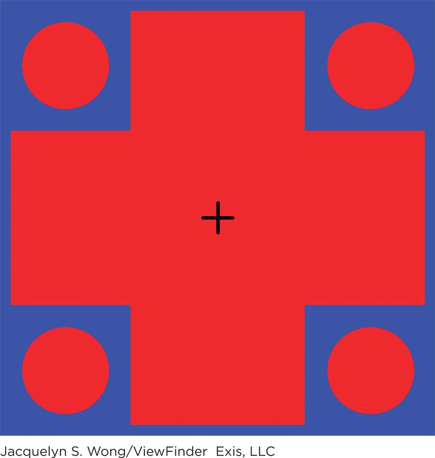
try this
Fix your eyes on the black cross in the center of
the image to the left. After 30 seconds, shift your gaze to
the blank white area. What colors do you see now?
Jacquelyn S. Wong/ViewFinder Exis, LLC
opponent-

A couple in China poses for a wedding photo. The bridal gown and roses are red, a color that connotes luck in Chinese culture. Many brides also wear red in India, where this color is associated with purity (Akcay, Dalgin, & Bhatnagar, 2011).
OPPONENT-
As it turns out, we need both the trichromatic and opponent-
Vision is a highly complex—
show what you know
Question 1
1. The hue of a color is determined by the ____________ of the light reflecting off an object.
wavelength
Question 2
2. Cells in the retina that absorb light energy and turn it into chemical and electrical signals are called ____________.
opponent-
processing photoreceptors
fovea
feature detectors
b. photoreceptors
Question 3
3. Explain the two major theories of color vision.
The trichromatic theory of color vision suggests there are three types of cones, each sensitive to particular wavelengths in the red, green, and blue spectrums. The brain identifies a precise hue by calculating patterns of excitement among the three types of cones, that is, the relative activity of the three types.
The opponent-
Question 4
4. It’s dark in your house, and you are struggling to see what time it is without turning on the light. You notice that if you turn your gaze slightly to the side of your watch, you can make out the large numbers. The ability to see these large details in the dark is due to your:
presbyopia.
optic disc.
cones.
rods.
d. rods.