3.4 Smell, Taste, Touch: The Chemical and Skin Senses
CONNECTIONS
In Chapter 2, we introduced the concept of neuroplasticity, which refers to the ability of the brain to heal, grow new connections, and make do with what’s available. Here, we suggest that Zoe’s heavy reliance on other senses is made possible by the plasticity of her nervous system.

Zoe rocks on a spring rider at the playground. The triplets spend much of their time outdoors—
 THE SCENT OF A MOTHER One day when Zoe was walking in the park with a friend, Liz decided to pay her an unexpected visit. As soon as her mom came within 15 feet, Zoe turned off the trail and made a beeline toward her with outstretched arms. She knew her mother had arrived because she could smell her, and she could do it while strolling through the breezy outdoors and eating a potent-
Smell: Nosing Around
LO 9 Describe the process of olfaction.
olfaction (ōl-
Many people take olfaction (ōl-
CONNECTIONS
In Chapter 2, we discussed the properties that allow neurons to communicate with other cells. When sending neurons signal a receiving neuron to pass along its message, their combined signal becomes excitatory and the neuron will fire. Here we are discussing olfactory receptor neurons stimulated by odor molecules. If enough odor molecules are present, an action potential occurs, causing a spike in electrical energy to pass through the axon.
A CHEMICAL SENSE Olfaction and taste are called chemical senses because they involve sensing chemicals in the environment. For olfaction, those chemicals are odor molecules riding currents of air. For taste, they are flavor molecules surfing on waves of saliva. Odor molecules, which are emitted by a variety of sources (for example, spices, fruits, flowers, bacteria, and skin), make their way into the nose by hitchhiking on currents of air flowing into the nostrils or through the mouth. About 3 inches into the nostrils is a patch of tissue called the olfactory epithelium. Around the size of a typical postage stamp, the olfactory epithelium is home to millions of olfactory receptor neurons that provide tiny docking sites, or receptors, for odor molecules (much as a lock acts as a docking site for a key; Figure 3.3 below). When enough odor molecules attach to an olfactory receptor neuron, it fires, causing an action potential, illustrating how transduction occurs in the chemical sense of olfaction.
OLFACTION IN THE BRAIN Olfactory receptor neurons stimulate a part of the brain called the olfactory bulb, where they converge in clusters called glomeruli (Figure 3.3). Then the signal is passed along to higher brain centers, including the hippocampus, amygdala, and olfactory cortex (Firestein, 2001). The other sensory systems relay data through the thalamus before going to higher brain centers. But the wiring of the olfactory system is unique; olfaction is on a fast track to the limbic system, where emotions like fear and anger are processed.
Humans have about 350 different types of odor receptors, yet we can distinguish over 1 trillion smells (Bushdid, Magnasco, Vosshall, & Keller, 2014). Each receptor type recognizes several odors, and each odor activates several receptors; a given scent creates a telltale pattern of electrical activity that the brain recognizes as lemon, garlic, or smelly feet. The types of receptors and the degree to which they are activated identify the odor (Firestein, 2001).
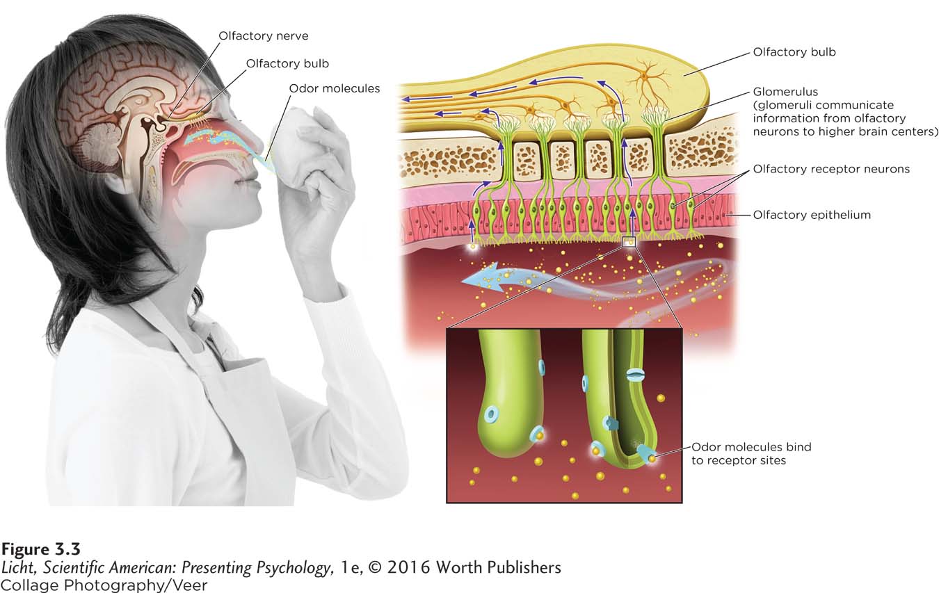
When enough odor molecules attach to an olfactory receptor neuron, it fires, sending a message to the olfactory bulb in the brain. Glomeruli then communicate the signal to higher brain centers.
Scents have a powerful influence on human behavior and thought. Minutes after birth, babies use the scent of their mother’s breast to guide them toward the nipple, and they quickly learn to discriminate Mom’s milk from someone else’s (Makin & Porter, 1989; Porter & Winberg, 1999). Research suggests that odor-
RECEPTOR REPLENISHMENT There is another reason smell is special. Olfactory receptor neurons are one of the few types of neurons that regenerate. Once an olfactory receptor neuron is born, it does its job for a minimum of 30 days, then dies and is replaced (Schiffman, 1997). This process slows with age, impairing odor sensitivity (Larsson, Finkel, & Pedersen, 2000). If it weren’t for this continual replenishment of olfactory neurons, we wouldn’t smell much of anything. Olfactory neurons are in direct contact with the environment, so they are constantly under assault by bacteria, dirt, and noxious chemicals. Some toxins—
Taste: Just Eat It
Eating is as much an olfactory experience as it is a taste experience. Just think back to the last time your nose was clogged from a really bad cold. How did your meals taste? When you chew, odors from food float up into your nose, creating a flavor that you perceive as “taste,” when it’s actually smell. If this mouth–
Tie on a blindfold, squeeze your nostrils, and ask a friend to hand you a wedge of apple and a wedge of onion, both on toothpicks so that you can’t feel their texture. Now bite into both. Without your nose, you probably can’t tell the difference (Rosenblum, 2010).
try this
LO 10 Discuss the structures involved in taste and describe how they work.
gustation (gəs-
ANOTHER CHEMICAL SENSE If the nose is so crucial for flavor appreciation, then what role does the mouth play? Receptors in the mouth are sensitive to five basic but very important tastes: sweet, salty, sour, bitter, and umami. You are likely familiar with all these tastes, except perhaps umami, which is a savory taste found in seaweed, aged cheeses, protein-
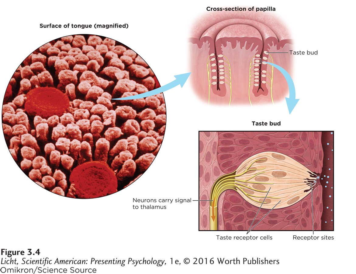
Taste buds located in the papillae are made up of receptor cells that communicate signals to the brain when stimulated by chemicals from food and other substances.
Receptor cells for taste are found in the taste buds on the tongue, the roof of the mouth, and lining the cheeks. Somewhere between 5,000 to 10,000 taste buds are embedded in the papillae, those bumps you can see on a person’s tongue (Society for Neuroscience, 2012). Jutting from each of these buds are 50 to 100 taste receptor cells, and it is onto these cells that food molecules bind (similar to the lock-
CONNECTIONS
In Chapter 2, we learned that sensory neurons receive information from the sensory system and send it to the brain for processing. With gustation, the sensory neurons are sending information about taste. Once processed in the brain, information can be sent back through motor neurons signaling you to take another bite!
As you bite into a juicy orange and begin to chew, chemicals from the orange (sour acid and sweet sugar) are released into your saliva, where they dissolve and bathe the taste buds throughout your mouth. The chemicals find their way to matching receptors and latch on, sparking action potentials in sensory neurons, another example of transduction. Signals are then sent through sensory neurons to the thalamus, and then on to higher brain centers for processing.
Receptors for taste are constantly being replenished, but their life span is only about 10 days (Schiffman, 1997). If they didn’t regenerate, you would be in trouble every time you burned your tongue sipping hot coffee or soup. Even so, by age 20, you have already lost half of the taste receptors you had at birth. And as the years go by, their turnover rate gets slower and slower, making it harder to appreciate the basic taste sensations (Kaneda et al., 2000). Drinking alcohol and smoking worsen the problem by impairing the ability of receptors to receive taste molecules. Losing taste is unfortunate, and it may take away from life’s simple pleasures, but it’s probably not going to kill you—
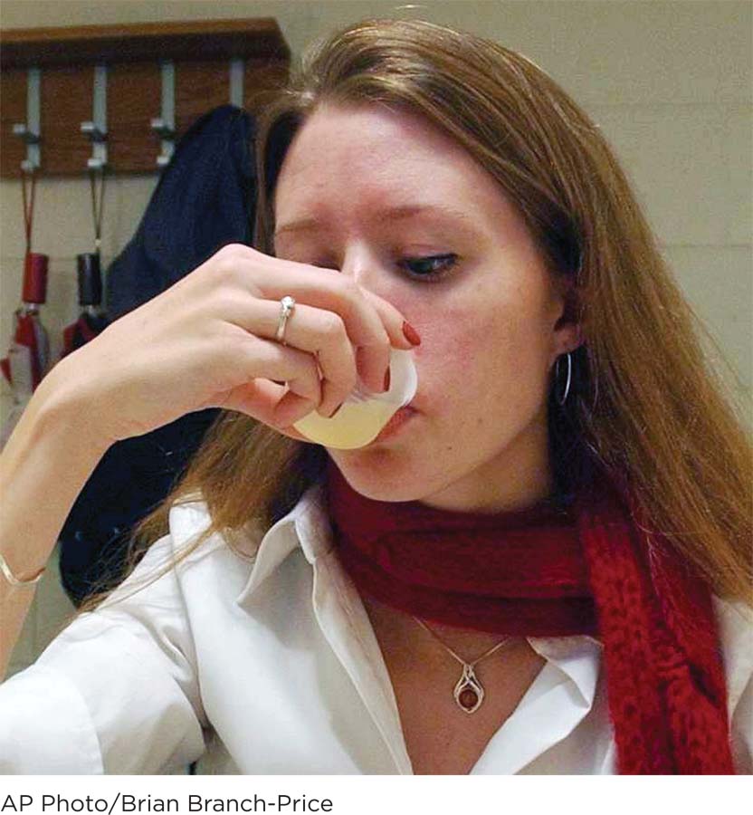
A graduate student performs a taste test at Rutgers University’s Sensory Evaluation Laboratory. Researchers have found that some people are more sensitive than others to bitter-
EVOLUTION AND TASTE The ability to taste has been essential to the survival of our species. Tastes push us toward foods we need and away from those that could harm us. We gravitate toward sweet, calorie-
Every person experiences taste in a unique way. You like cilantro; your friend thinks it tastes like bath soap. Taste preferences may begin developing before birth, as flavors consumed by a pregnant woman pass into amniotic fluid and are swallowed by the fetus. In one study, infants who had been exposed to the flavor of carrots before birth (through their mothers’ consumption of carrot juice in the last trimester) showed fewer disapproving facial expressions when fed carrot-
We have made some major headway in this chapter, examining four of the major sensory systems: vision, hearing, smell, and taste. Now it is time to get a feel for a fifth sense. Are you ready for tingling, tickling, and titillating touch?
Touch: Feel the Magic

Zoe approaches eating in a very tactile way, digging in and feeling the food on her skin. While Zoe cannot see or hear, her senses of smell, taste, and touch are extremely fine-
If you were introduced to Zoe, she would probably explore your face for about 20 seconds, and then return to whatever she was doing. Emma would likely spend more time getting to know you. She might give you a hug, put her cheek against yours, and carefully examine your head with her fingers. At the dinner table, Emma is neat and tidy, and does not like to get food on her hands and face. Zoe loves to dig in and make a mess. These two girls, who are genetically identical, experience the world of touch in dramatically different ways. But both rely on touch receptors within the body’s main touch organ—
Take a moment to appreciate your vast epidermis, the outer layer of your skin. Weighing around 6 pounds on the average adult, the skin is the body’s biggest organ and the barrier that protects our insides from cruel elements of the environment (bacteria, viruses, and physical objects) and distinguishes us from others (fingerprints, birthmarks). It also shields us from the cold, sweats to cool us down, and makes vitamin D (Bikle, 2004). And perhaps most importantly, skin is a data collector. Every moment of the day, receptors in our skin gather information about the environment. Among them are thermoreceptors that sense hot or cold, Pacinian corpuscles that detect vibrations, and Meissner’s corpuscles sensitive to the slightest touch, like a snowflake landing on your nose (Bandell, Macpherson, & Patapoutian, 2007; Figure 3.5).
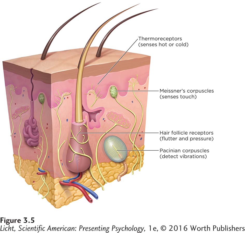
The sensation of touch begins with our skin, which houses a variety of receptors including those shown here.
Oh, the Pain
Not all touch sensations are as pleasant as the tickle of a snowflake. Touch receptors can also communicate signals that are interpreted as pain. Nociceptive pain is caused by heat, cold, chemicals, and pressure. Nociceptors that respond to these stimuli are primarily housed in the skin, but they also are found in muscles and internal organs.
In very rare cases, a baby is born without the ability to feel pain. Children with this type of genetic disorder may face a greater risk of serious injury (Romero, Simón, Garcia-
CONNECTIONS
In Chapter 2, we explained how a myelin sheath insulates the axon and speeds up the conduction of electrical impulses. The myelin covering the fast nerve fibers allows them to convey their information about pain more quickly than the slow nerve fibers that are unmyelinated.
TWO PATHWAYS FOR PAIN Thanks to the elaborate system of nerves running up and down our bodies, we are able to experience unpleasant, yet very necessary, sensations of pain. Fast nerve fibers, made up of large, myelinated neurons, are responsible for conveying information about pain occurring in the skin and muscles, generally experienced as a stinging feeling in a specific location. If you stub your toe, your first perception is a painful sting where the impact occurred. Slow nerve fibers, made up of smaller, unmyelinated neurons, are responsible for conveying information about pain throughout the body. Pain conveyed by the slow nerve fibers is more like a dull ache, not necessarily concentrated in a specific region. The diffuse aching sensation that follows the initial sting of the stubbed toe results from activity in the slow nerve fibers. As the names suggest, fast nerve fibers convey information quickly (at a speed of 30–
The axons of these fast and slow sensory nerve fibers band together as nerves on their way to the spinal cord and up to the brain (Figure 3.6). The bundled fast nerve fibers make their way to the reticular formation of the brain, alerting it that something important has happened. The information then goes to the thalamus and on to the somatosensory cortex, where sensory information from the skin is processed further (for example, indicating where it hurts most). The bundled slow nerve fibers start out in the same direction, toward the brain, with processing occurring in the brainstem, hypothalamus, thalamus, and limbic system. In the midbrain and amygdala, for example, emotional reactions to the pain are processed (Gatchel, Peng, Peters, Fuchs, & Turk, 2007). As with other neurons, the transmission of information, in this case about pain, occurs through electrical and chemical activities. Substance P and glutamate are two important neurotransmitters that work together to increase the firing of the pain fibers at the injury location.
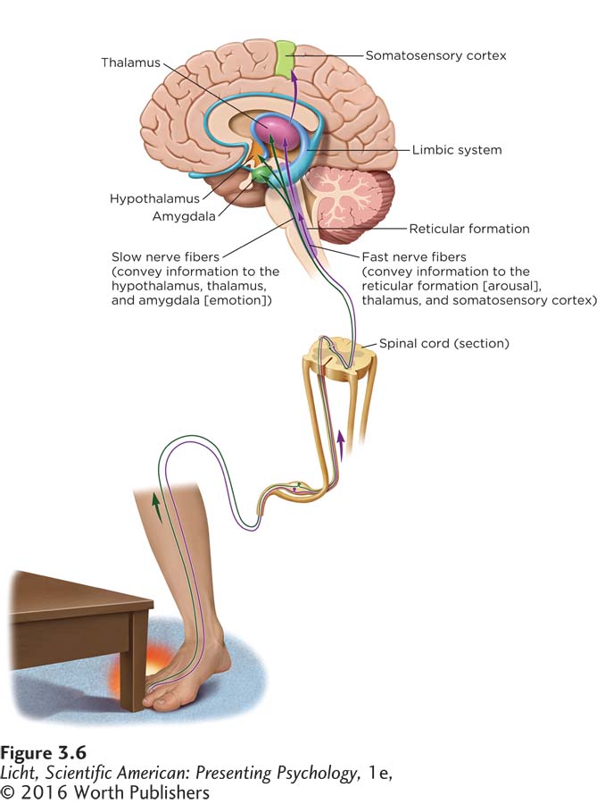
When you stub your toe, two kinds of pain messages can be communicated to your brain. Your first perception of pain may be a sharp, clear feeling where the impact occurred. The message quickly travels through your spinal cord to your brain, signaling arousal and alerting you to react. Slow nerve fibers also travel through your spinal cord to carry messages about the pain that lingers after the initial injury, often generating an emotional response.
Understanding the mechanisms of pain at the neural level is important, but biology alone cannot explain how we perceive pain and why people experience it so differently. How can the same flu shot cause great pain in one person but mere discomfort in another? And why does our sensitivity to pain seem to fluctuate from one day to the next?
LO 11 Describe how the biopsychosocial perspective is used to understand pain.
As with most complex topics in psychology, pain is best understood using a multilevel method, such as the biopsychosocial perspective. Chronic pain, for example, can be explained by biological factors (the neurological pathways involved), psychological factors (distress, cognition), and social factors (immediate environment, relationships; Gatchel et al., 2007). Prior experiences, environmental factors, and cultural expectations influence how the pain is processed (Gatchell & Maddrey, 2004).
Researchers studying pain once searched for a direct path from pain receptors to specific locations in the brain, but this simple relationship does not exist (Melzack, 2008). Instead, there is a complex interaction between neurological pathways and psychological and social factors, and gates involved in the shuttling of information back and forth between the brain and the rest of the body.
GATE CONTROL The most influential theory of pain perception is the gate-
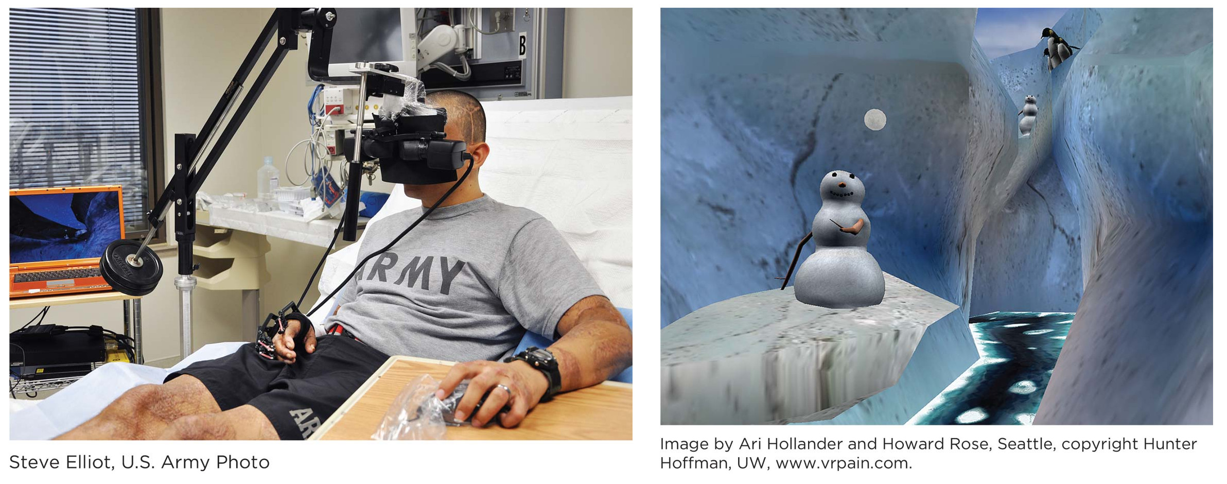
The last thing you want to look at while undergoing an unpleasant or painful medical procedure is the ceiling tiles in your hospital room. Being immersed in a virtual winter scene, complete with snowmen and penguins, can help take one’s mind off the pain (Li, Montaño, Chen, & Gold, 2011). Army Sergeant Oscar Liberato (left) gets lost in the virtual reality video game SnowWorld (right) as he undergoes a painful procedure.
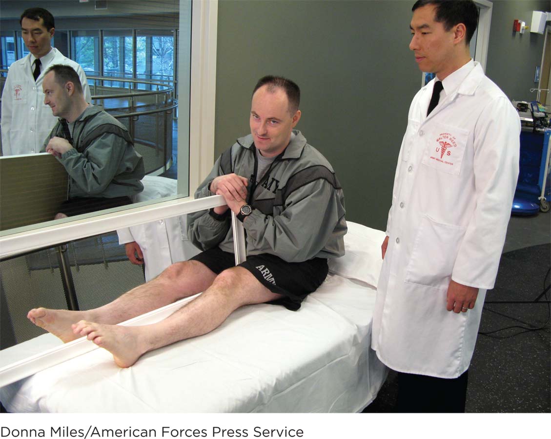
Neurologist Jack Tsao uses mirror therapy to help Army Sergeant Nicholas Paupore at the Walter Reed Army Medical Center. The reflection of a leg in the mirror seems to take on the identity of the one that is missing, which may help the patient resolve his phantom limb pain. Mirror therapy is a promising development, but further studies are needed to evaluate its efficacy (Moseley, Gallace, & Spence, 2008).
Signals to shut the gate do not always come from the brain (Melzack, 1993, 2008). Information sent from the pain receptors in the body can also open and close the neurological gates from the bottom up, depending on which type of fiber is active. If the small unmyelinated fibers are active, the gates are more likely to open, sending the pain message up the spinal cord to the brain. If the large myelinated fibers are active, the gates are more likely to close, inhibiting pain messages from being sent on. Suppose you stub your toe. One way to ease the pain is by rubbing the injured area, stimulating the pressure receptors of the large fibers. This activity closes the gates, interfering with the pain message that would otherwise be sent to the brain.
It is clear that everyone experiences pain in a unique way. But are there factors that predispose a person to be more or less sensitive to pain?
CONNECTIONS
In Chapter 2, we reported that endorphins are naturally produced opioids that regulate the secretion of other neurotransmitters.
THE PSYCHOLOGY OF PAIN Pain sensitivity can ebb and flow for an individual, with psychological factors exerting a powerful effect (Gatchel et al., 2007; Raichle, Hanley, Jensen, & Cardenas, 2007). Negative feelings such as fear and anxiety can amplify pain, whereas laughter and distraction can soften it, interfering with the ability to attend to pain. Certain types of stressors (for example, running in a marathon, or giving birth) trigger the release of endorphins. Endorphins reduce pain by blocking the transmission of pain signals to the brain and spinal cord, possibly through the inhibition of substance P, a neurotransmitter in the spinal cord and brain (Rosenkranz, 2007).
PHANTOM LIMB PAIN Perhaps the most striking illustration of pain’s complexity is phantom limb pain. Some 50–
Kinesthesia: Body Position and Movement
LO 12 Illustrate how we sense the position and movement of our bodies.
kinesthesia (ki-
proprioceptors (proh-

Without his sense of kinesthesia, stuntman Nik Wallenda never would have crossed the Niagara Falls on an 1,800-
You may have thought “touch” was the fifth and final sense, but there is more to sensation and perception than the traditional five categories. Closely related to touch is the sense of kinesthesia (ki-
vestibular sense (ve-
You may recall from earlier in the chapter that Emma, Zoe, and Sophie began to curl up in the fetal position and vomit while riding in the car during the same period that they lost their hearing. This was because they were losing their vestibular sense (ve-
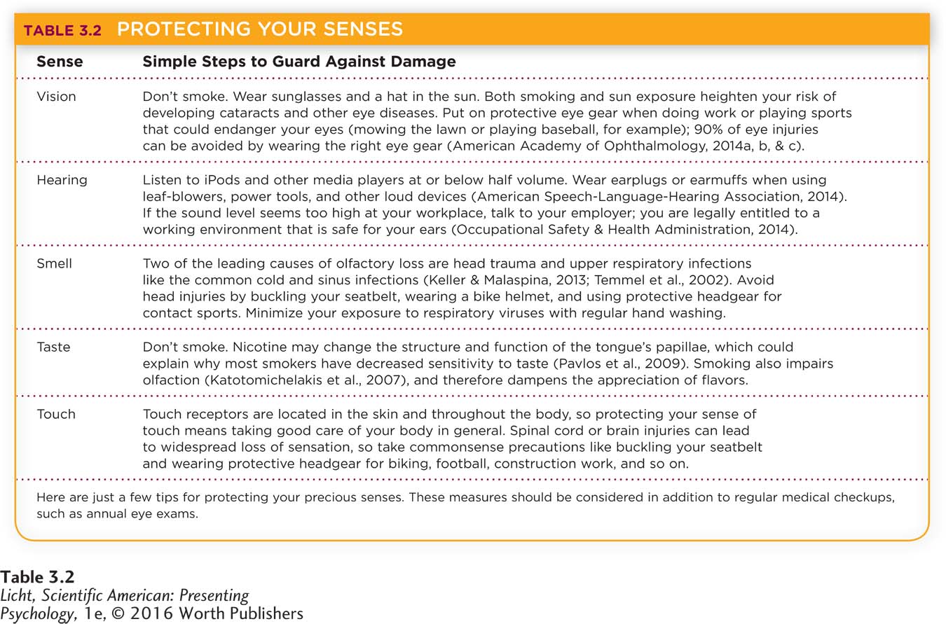
Before we move on to the next section on perception, let’s take a moment to appreciate the sensory systems that allow us to know and adapt to the surrounding world. How would you function without vision, hearing, smell, taste, and touch, and what steps can you take to preserve them (Table 3.2)?
Apply This
show what you know
Question 1
1. The chemical sense called ____________ provides the sensation of smell.
olfaction
Question 2
2. Chemicals from food are released in saliva, where they dissolve and bathe the taste buds. The chemicals find matching receptors and latch on, sparking action potentials. This is an example of:
olfaction.
transduction.
sensory adaptation.
thermoreceptors.
b. transduction.
Question 3
3. List five things you are currently doing that involve the use of kinesthesia.
Answers will vary. Some examples may include typing on your computer, sitting in your chair, getting food from the refrigerator, turning off the fan, taking notes.
Question 4
4. Maya consulted her physician about severe back pain. In order to help her understand pain perception, the doctor recommended she consider ____________, which suggests that a variety of biopsychosocial factors can interact to amplify or diminish pain perception.
the theory of evolution
an absolute threshold
the gate-
control theory gustation
c. the gate-