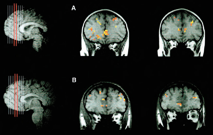
fMRI images The figure shows fMRI images of the brains of a 9-year-old (panel A) and a 24-year-old (panel B) in a standard cognitive task that requires responding to some stimuli but inhibiting responding to others. (The images to the left show the “slices” of the brain averaged together in the images on the right.) The results show that the location of activation in the prefrontal cortex did not differ between children and adults, but the overall extent of activation was greater for the children (Casey, 1999).
REPRINTED WITH PERMISSION FROM B. J. CASEY (1999)