Chapter 10. ANIMAL DIVERSITY 2
I. OVERVIEW
Last week we started looking at the diversity of the Animal Kingdom and at the various ways that animals are grouped. We started with the Porifera, a group that lacks true tissues but does have specialized cells, and has an important place in marine ecosystems. We then looked at Cnidaria, a phylum that shows radial symmetry, is diploblastic, has specialized cells, and lacks cephalization. Members of the Cnidaria often show an alternation of forms (polyp and medusa) and are very important in marine ecosystems.
Next, we looked at examples that show bilateral symmetry and at least some degree of cephalization. The Protostomata include the Lophotrochozoa and the Ecdysozoa. Last week we looked at the three phyla in the Lophotrochozoa, the Platyhelminthes (including the Dugesia and the important parasites: the flukes and tapeworms), the Phylum Annelida (characterized by their worm shape and segmentation), and the Phylum Mollusca (important and complex examples include the snails, bivalves, and cephalopods).
In this lab, we will continue our comparison of animals by looking at the other group within the protostomes, the Ecdysozoa. And we will finish by looking at the deuterostomes. You can learn a lot more about all of the groups covered in these labs by taking the Invertebrate Zoology class, offered by Dr. Gaston. In addition, biology classes offered in Belize and in the Galapagos give you opportunities to study many of these groups. The Biology Department offers a class in the Biology of Fishes by Dr. Parsons and a Mammalogy class, offered by Dr. Buchholz.
II. THE ECDYSOZOA
In the Ecdysozoa, we will look at the Phyla Nematoda and Arthropoda. Ecdysozoa include the two phyla that grow through a process called ecdysis or molting. That is, members of this group grow by shedding their skins. This relationship is supported by a comparison of many genes, including ribosomal RNA genes. This is a good example when there are phyla that do not necessarily look very much alike (e.g., roundworms and insects) yet we now know that they share important characteristics and a common ancestry. Nematodes evolved a pseudocoelom through simplification of a more complex body plan.
PHYLUM NEMATODA
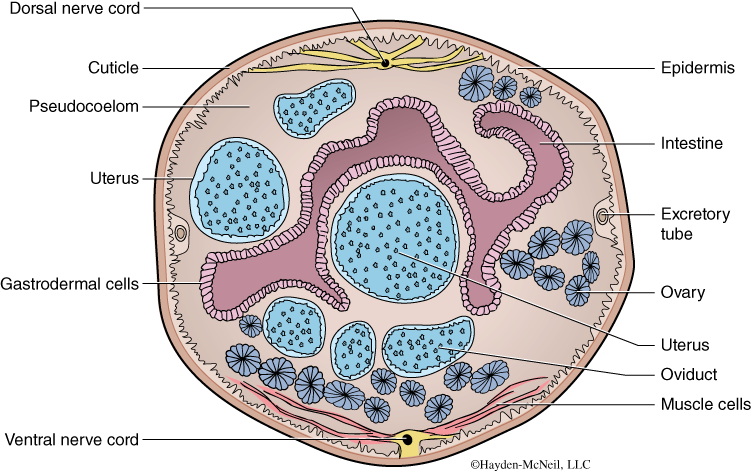
The phylum Nematoda are also called the roundworms because they are round in cross section (compared to the flatworms, studied last week). In addition, compared to the Annelida (also studied last week), they have a relatively simple internal structure (e.g., see Figs. 10.1 and 10.2). Many nematodes are parasites and they vary greatly in size. Roundworms are common and abundant but often overlooked because many of them are small in size. It has been said that if the trees or soil disappeared but the nematodes remained, you would still see an outline of everything. They are very conservative in shape; that is, they all look very similar and it is difficult to tell one species from another. They have bilateral symmetry, a tube-within-a-tube digestive system, and like many parasites, have impressive abilities to reproduce. They have a body cavity called a pseudocoelom, which is a fluid-filled cavity lined by tissue that is not entirely from mesoderm. We will look at vinegar eels and the Ascaris roundworm. They have longitudinal muscles and a hydrostatic skeleton and can only move by whipping back and forth. Thus, they are not able to move quickly but can probably move efficiently in a medium such as soil or in the gut of another animal.
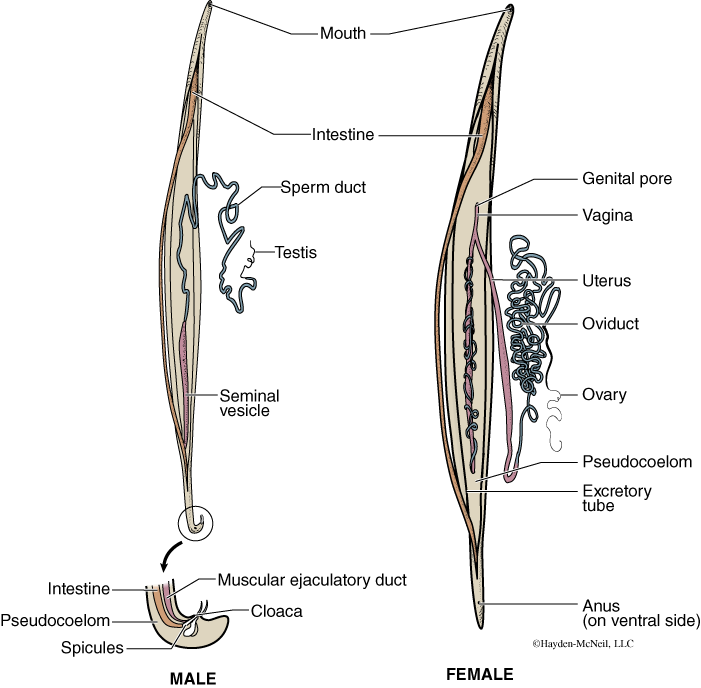
The Ascaris demonstrates nicely how a parasitic animal will devote most of its body mass to reproduction. They are dieocious (sexes are separate), so each table as a group will look at both a dissected male and a female. Your text is helpful for providing information about other important nematode parasites.
PROCEDURE
Examine the live vinegar eels, a type of nematode, under the dissecting scope and pay particular attention to how they move. Note that they can only whip back and forth, not lengthen and shorten like the earthworm. Next, examine in detail the dissected male and female worms that are on each table. The first lab section will do the dissection. Sketch the worms and identify the parts listed in Fig. 10.2. Finally, examine the cross section of the worms with a compound microscope (Fig. 10.1) . On the female cross section, identify the ovary and the uterus. Why do these worms need to make so many offspring? What are the differences between the ovaries, the oviducts, and the uterus? On your sketch of the whole female worm, indicate from where this cross section was taken.
PHYLUM ARTHROPODA
The Phylum Arthropoda is the most successful and largest of all animal phyla. It is a phylum comprised of more than a million species and includes well-known examples (lobsters, shrimp, insects, and spiders) and lesser-known examples (such as amblypigids and isopods). Nearly 80% of the Animal Kingdom is in this phylum. Why are arthropods so successful? Arthropods have a clearly segmented body. In evolution, this segmentation has allowed for certain regions of the body (known as tagmata) to be specialized for particular functions. For example, the head region often has a concentration of sensory organs and the thorax is specialized for locomotion. Arthropods have an exoskeleton make of chitin. This protects these animals from desiccation or drying out and also serves as a protection from predators. In addition, arthropods have paired jointed appendages. These appendages are also specialized, even within a single species, as we will see in the crayfish. For example, some legs are specializing for walking, some for sensing the environment, and some may be specialized for swimming. In addition, arthropods are relatively small, often have a relatively short generation time and in many cases can produce a large number of offspring. All of these characteristics have contributed to their evolutionary success.
Here is the taxonomy that you are responsible for in this class. For each of these, you need to be able to recognize the examples that will be displayed in lab. For each, note if there are distinct regions of the body, note the number and specialization of legs and the presence or absence of wings.
Arthropoda
Myriapoda: Millipedes and centipedes
Arachnida: Spiders, ticks, scorpions
Insecta: Beetles, wasps, flies
The University of Mississippi Biology Department has several people who work with arthropods. Dr. Paul Lago is a world-class entomologist who specializes in beetles. People from all over the world send him specimens to identify. Dr. Gail Stratton is an arachnologist with an interest in the behavior, ecology, and systematics of wolf spiders.
Our goals in working with Arthropoda in this lab are to first look at the external morphology of one group as a representative of this large and distinctive phylum. Second, we will look at examples of the main groups.
The Crustacea
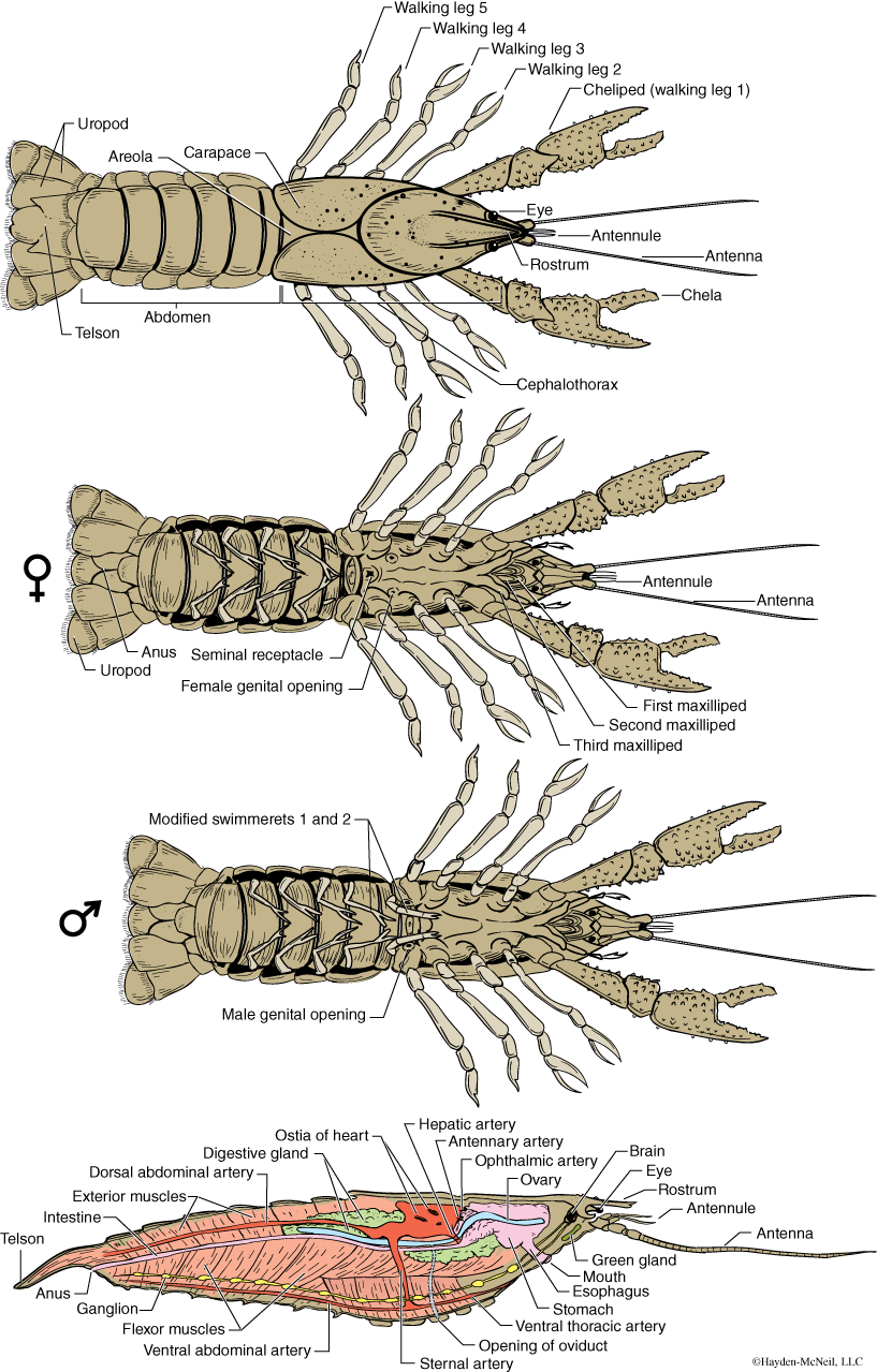
We will start with a detailed look at a common member of the crustacea, the crayfish (Fig. 10.3). The midsouth region of the United States and Mississippi in particular has a remarkable diversity of crayfish. Crayfish live in freshwater and are able to move both forward and backward. They use their powerful abdominal muscles for quickly moving backward into hiding places. Their large chelipeds are used for defense. They are scavengers.
The Myriapoda
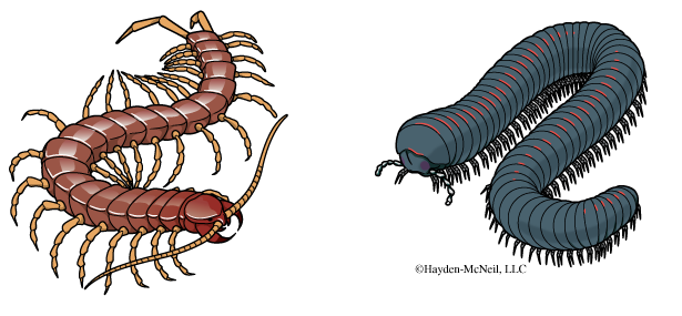
The best-known Myriapoda are the centipedes and millipedes. These elongate terrestrial arthropods show very little specialization of regions (that is, the front end looks very much like the hind end). Centipedes have one pair of walking legs on each segment of their bodies, while millipedes have two pairs on each segment. Millipedes tend to be round in cross section and are herbivores and scavengers. Centipedes are much flatter in cross section and are predators with a poison fang near their mouth. Which one should you be careful picking up?
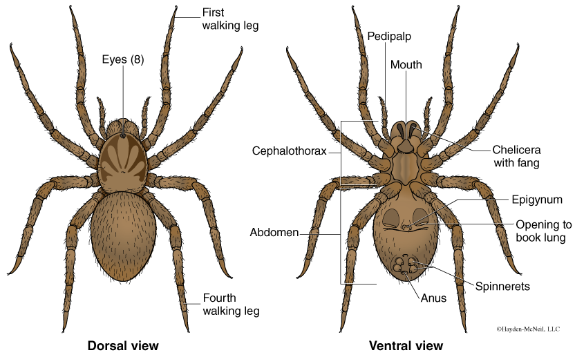
Class Arachnida
Arachnids include a fascinating variety of animals including spiders, ticks and scorpions. There are about 30,000 named species of spiders and many more that are not yet given a formal name. In Mississippi, there are around 600 different species of spiders; of the 600, only three species are potentially hazardous to people (there are two species of black widows and one species of brown recluse). We will have several examples of spiders available in lab, including the brown recluse and black widows, spiders everyone should be able to recognize.
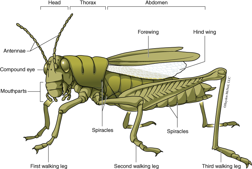
Class Insecta
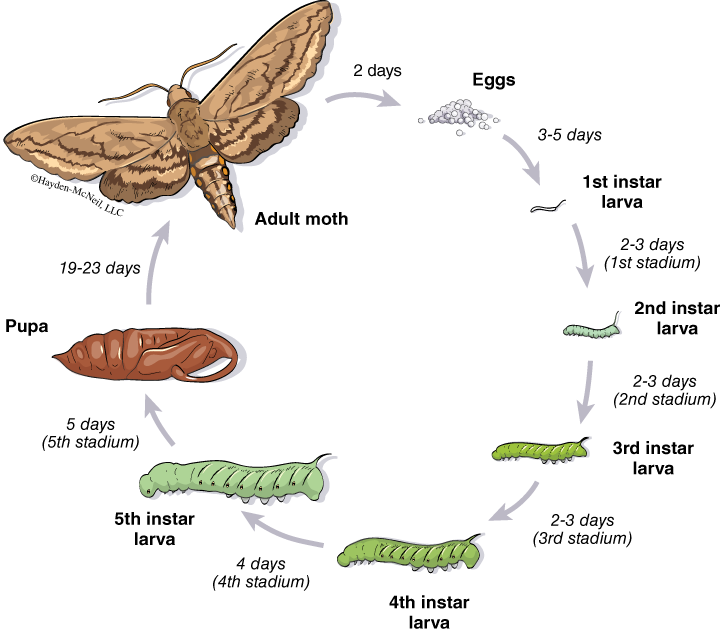
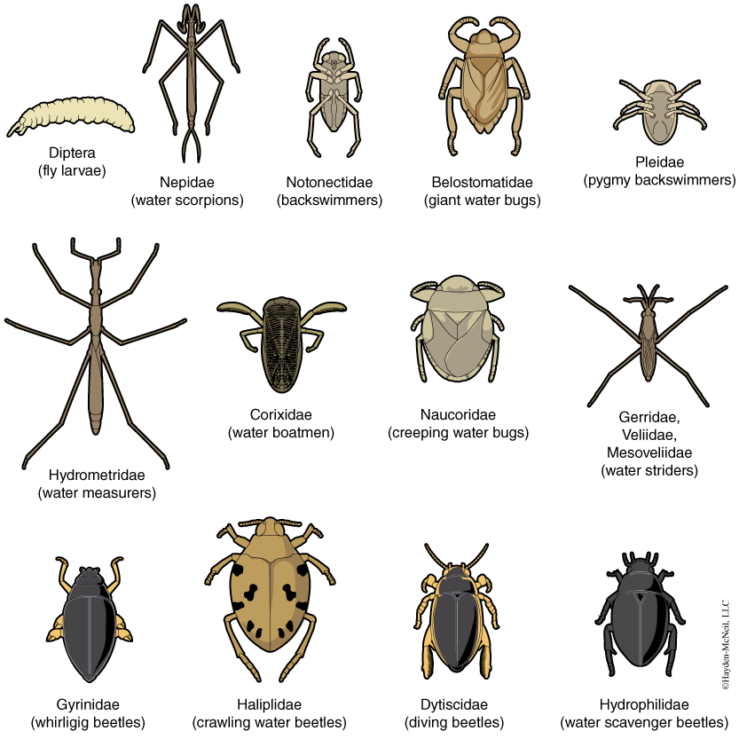
The Insecta is the most successful group of animals on the planet. Dr. Lago’s entomology class is an opportunity to study them in more detail than we can here. Although not all insects have wings, many of the largest groups of insects do have wings, and it is thought that the ability to disperse readily was very important in the evolution of their diversity. In lab we will have examples out of some of the more common groups of insects: grasshoppers (Fig.10.6), beetles, flies, and wasps. Many insects have a complete metamorphosis, in which the larval stage looks completely different from the adult (Fig. 10.7). Fig. 10.8 shows a glimpse at some of the diversity within this large and important class.
PROCEDURE
We will start with the external morphology of a crayfish. Look at the crayfish on the lab tables. First look at the animal dorsally and identify these tagmata: cephalothorax, abdomen, and tail. Find the eyes, antennae, carapace, cheliped, and walking legs. Make a sketch with these structures labeled. Handle the specimens gently!
Next, examine the ventral surface of the crayfish and find these modified appendages: the uropod (literally “tail foot”), the swimmerets or pleopods, the walking legs, the cheliped, and the mouth appendages. From looking at these appendages, you should now have a sense of how arthropods, even in one individual, can use the specialization of appendages in different regions of their bodies to great advantage in evolution. That is, through evolution, a jointed appendage can be modified to a sensory laden mouth part, or be modified with pincers for picking up food, or can be broadened and be part of a tail fan. Finally, you should determine if your crayfish is a male or female. Look around the room at other crayfish and be sure the see an example of each sex.
Compare the other subphyla of arthropods. Along the side table are examples of each of the subphyla. For each, you should be able identify phylum, subphylum, class, and common names. Note if there are distinct body regions, number and specialization of legs, and presence or lack of wings. Make a table that summarizes this comparison.
III. THE DEUTEROSTOMATA
The final large group of animals that we will consider are the deuterostomes. There are two phyla in the deuterostomes that we will look at in some detail: the Echinodermata and the Chordata. The Chordata include the Subphylum Vertebrata and thus includes most of the animals with which we are familiar (mammals, birds, reptiles and fish). Although the echinoderms and chordates look very different (for example, compare a starfish and a horse!), both phyla are united by their similarity in early development and also because of similarities in the sequences of many genes. You recall that deuterostomes have radial, indeterminate cleavage, and that the mouth does not develop from the blastopore. By this point, you should be very comfortable with these terms.
PHYLUM ECHINODERMATA
This phylum is an ancient group, with representatives from the very earliest multicellular animals, 600 million years ago. Because they have an endoskeleton of calcium carbonate, which fossilizes readily, there in an excellent fossil record of this phylum. In fact, during the Paleozoic Era (248–570 million years before present) there were more classes or major groups of echinoderms than there are now. At one time there were 20 different classes in the phylum; now there are five. We will examine examples from several different classes in this lab. And, although they are an ancient group, they have a highly derived body plan that is quite different from anything else we will see.
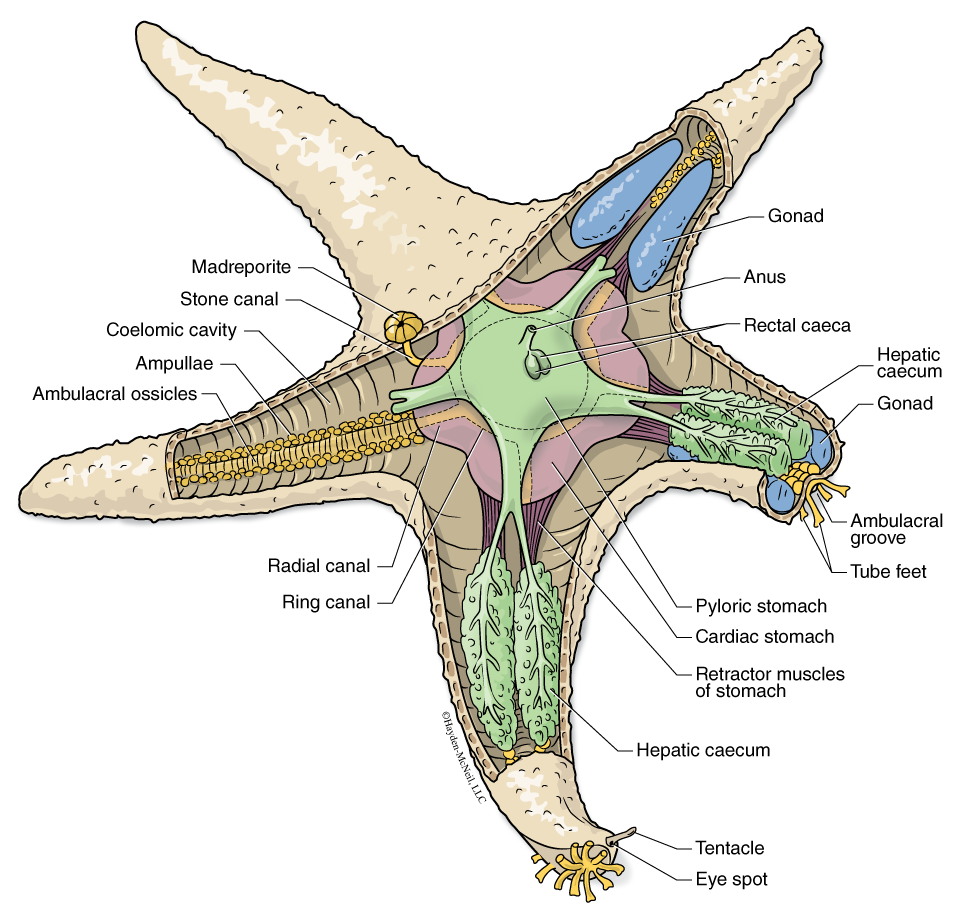
The larvae of this phylum are bilaterally symmetrical and are free swimming and quite mobile, even though they are small. It is the larval stage that disperses. In the process of maturation, they go through a drastic transformation (a metamorphosis) and become much less mobile and they reorganize to have a pentaradial symmetry or a five-part symmetry around a radial disk. Uniquely in the animal kingdom, they have a system called the water vascular system, a system of fluid-filled tubes canals and chambers that simultaneously functions a locomotor system (via the tube feet), circulatory system, and respiratory system. One of the best places to see this system is in the sea stars, or Asteroidea (also called the starfish). We will look at preserved specimens. In addition, there is a live sea star in the large marine tank in the entryway of Shoemaker.
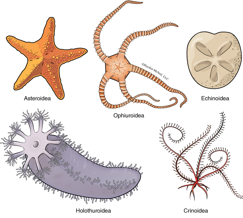
Here is the taxonomy of Echinodermata for which you are responsible (Fig. 10.10).
Echinoidea, sea urchins and sand dollars
Holothuroidea, sea cucumbers
Ophiuroidea, brittle stars
Crinoidea, feather stars
PROCEDURE
Each group of two people should get a prepared slide of early sea star development and on the slide, find and sketch the larval stage.
Each group of two students will have a preserved sea star, a member of the Class Asteroidea. One example will be dissected so you can see the internal aspect of the water vascular system. On these animals, you should find, identify, and sketch these structures: tube feet, radial disk and the madreporite, a possible valve or opening in the water vascular system. If the animal is mature, you may see gonads or the digestive system in the legs. Your sketch should be labeled.
Next you need to look at, sketch and know the other examples on display: the Holothuroidea, or sea cucumbers; the Echinoidea, or sea urchins and sand dollar; the Ophiuroidea, or brittle stars; and the Crinoidea. Finally, you should look at the cross section of the arm of a starfish, to see how the water vascular system is suspended in the arm. Complete a table that summarizes each of the classes, the common name and a trait that distinguishes that class.
PHYLUM CHORDATA
The Chordata include us, the humans, a species having the largest impact of any species in history on planet earth. Chordata is a large, important phylum and it includes some invertebrate members. There are three subphyla we will be examining and you need to know examples of each. The subphyla include the Urochordata (or sea squirts), the Cephalochordata (lancelets), and the best known of the three, the Vertebrata.
What characteristics unite these three subphyla? First, all chordates have a notochord some time in their development. A notochord is a dorsal stiff rod that helps to provide support to the animal. It also secretes proteins that are important in development. In vertebrates the notochord is replaced by the vertebral column, but remnants can remain in the first years of life. Next, all members have a dorsal, hollow nerve tube. All have pharyngeal gill slits at some time in their development; all have a post anal tail, again at some time in their development; and finally, all have an endostyle, a ciliated groove on the ventral wall of the pharynx. In tunicates and lancelets, this grove secretes mucus that helps the animal to catch food; in vertebrates, the endostyle develops into the thyroid gland, a secreting organ that is part of the endocrine system.
Here is the taxonomy of Chordata for which you are responsible:
Cephalochordata
Vertebrata
Urochordata
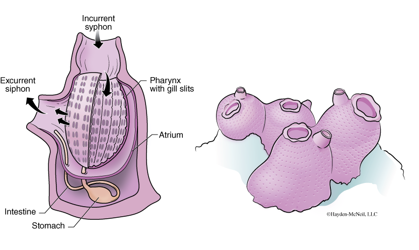
The tunicates are members of this group. They are sometimes call “sea squirts” because when they are touched, they “close down” by squirting all of the water in the siphons out quickly, They typically have two siphons (Fig. 10.11): an incurrent siphon and an excurrent siphon, and pull water in via the incurrent siphon to pass across a large structure that is a modified pharynx. It is modified by having many openings and cells that secrete a large amount of mucus. The mucus catches particles of food as the water moves across this structure and the food-laden mucus is then carried into the esophagus and is eaten.
Externally, the tunicates do not look like close relatives of the vertebrates, or even of the cephalochordates. Curiously, however, about 80% of the genes in at least some tunicates are shared with vertebrates, suggesting a very close relationship. The tunicate larva looks a lot like the Cephalochordata.
Subphylum Cephalochordata
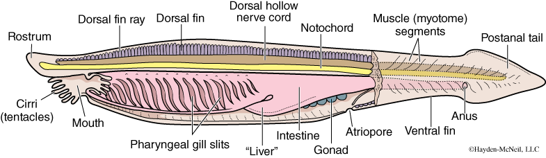
The common name for this group are the lancelets, or members of the genus Amphioxus or Brachiostoma (Fig. 10.12). These are curious animals that are shaped like a fish and live in shallow water in marine systems. They often burrow and feed in an manner that is similar to the Urochordata: they pull water across the pharynx, filter the water, and catch particles via the mucus. They have a clear notochord.
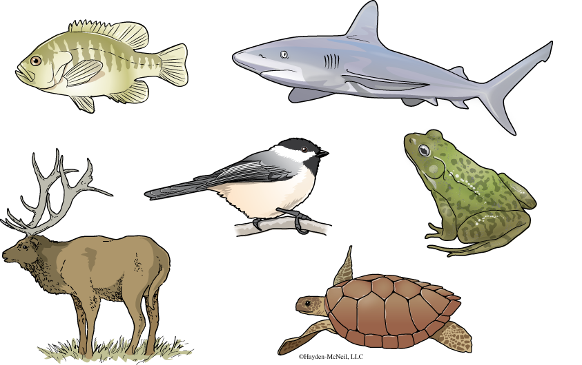
VERTEBRATA
What is a typical vertebrate? Unlike the first two subphyla in the Phylum Chordata, there is no single typical vertebrate. It is a large and varied subphylum; it is also a very old subphylum. The oldest possible representative looked like an elongate tadpole and lived 560 million years ago (Fig. 10.13).
Vertebrata (There are six separate classes of fish and four classes of tetrapods. We will cover only three of the fish classes.)
Actinopterygii
Actinistia
Dipnoi
Amphibia
Reptila
Aves
Mammalia
Chondrichthyes
These are the cartilaginous fish: the sharks, rays, and skates. The Ole Miss Biology Department is lucky to have on the of the premier shark experts on its faculty. Dr. Glenn Parsons. Dr. Parsons wrote the book, Sharks, Skates, and Rays of the Gulf of Mexico: A Field Guide.
Class Actinopterygii
These are the ray-finned fishes and include the fishes we are generally most familiar with: the perch, salmon, tuna, and trout. The are bony, have a swimbladder, and are in both marine and fresh water.
Class Actinistia
These are the lobe-finned fishes and include the so-called “living fossil,” the Coelacanth.
Class Dipnoi
These are lungfishes, bony freshwater fishes that have limbs.
Class Amphibia
The Class Amphibia includes caecilians, salamanders, toads and frogs. Typically, the larval stage is aquatic and the adult stage is terrestrial. They have a three-chambered heart, with two atria and one ventricle.
Class Reptilia
The Class Reptilia includes turtles, lizards, snakes, and alligators. It is considered a category of convenience and is a paraphyletic group. Members are amniotes with horny scales.
Class Aves
Class Aves, the birds, are an amazing group of animals that are in the same lineage as many reptiles. They are amniotes with feathers. They have wings (modified anterior limbs), a fourchambered heart, and are endothermic.
Class Mammalia
The Class Mammalia includes monotremes (such as the duck-billed platypus), marsupials (which includes kangaroos and many others), and the placental mammals, or those that have a placenta. Members of the class are amniotes, have hair and females feed their young with mammary glands. They have a four-chambered heart and are endothermic.
PROCEDURE
In this portion of the lab will start with the examination of the Urochordata and Cephalochordata. Look at preserved examples and pictures of the sea squirts (subphylum Urochordata) and preserved examples of the in the subphylum Cephalochordata. Use the dissecting scope for the latter. In addition, we have mounted examples of Amphioxus as well as cross sections of them. For the Amphioxus, you should make a sketch of it based on the mounted slide and label as many parts as you can discern (from Fig. 10.12).
Why are vertebrates grouped with the urochordates and the cephalochordates? Next, survey the examples of vertebrates that we have available and learn the class and common names. Be able to recognize each.
Make a table that summarizes the characteristics that each of these groups and the characteristics that make them unique. Refer to the phylogeny presented in Chapter 10.
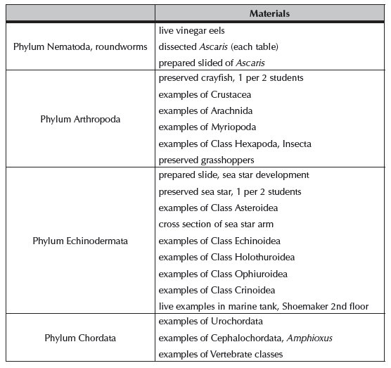
IV. NOTES TO INSTRUCTORS AND MATERIALS FOR LAB
To do well with this material, students must come to class with the major categories of animals clearly in mind.
Suggested time line for class:
30 min. quiz and introduction
30 min. Arthropoda
15 min. Chordates
20 min. Nematoda
30 min. Echinoderms
30 min. vertebrates
There will be material representing some of the diversity of these groups along the side tables in the lab. In addition, each student table should have two dissecting trays, each with this set of organisms:
- Good resource: http://www.pbs.org/kcet/shapeoflife/explorations/index.html
- All microscope slides have been sorted and labeled. It is everyone’s responsibility to be sure that slides are returned to the correct tray. Reminder concerning slides: W.M. = whole mount; C.S. = cross section