Chapter 3. EXTERNAL ANATOMY, DIGESTIVE SYSTEM, AND RESPIRATORY SYSTEM
I. OVERVIEW
Today we begin the close work with fetal pigs. Fetal pigs have often been used as model vertebrates for study in labs such as ours. And, since pigs and humans are both mammals, there are many ways we are anatomically similar. Throughout these labs we will be making comparisons of the fetal pig with other animals.
Pigs are members of the Kingdom Animalia, Phylum Chordata, Subphylum Vertebrata, Class Mammalia. Within the Class Mammalia, they are placed in the Artiodactyla, a group that is closely related to the Cetacea (the whales, dolphins, and porpoises). The Artiodactyla include the pigs, deer, cattle, sheep, goats, and camels, among others. Artiodactyla are called the “even-toed ungulates.” They have a cloven hoof with two weight-bearing toes, comprised of the third and fourth digits (digits 2 and 5 are reduced, the thumb is absent). Recent work has included the cetaceans in the same order as the Artiodactyla.
Pigs are in the Family Suidae, a family with 18 living species found all over the world. The domestic pig is the Sus scrofa. Pigs were domesticated from the Eurasian wild boar. There is genetic evidence that domestication occurred independently several times both in Europe and in Asia. Domestication occurred around 9,000 years ago.
In many areas, pigs have escaped from human control and are now feral. When this happens, they can become ecologically very destructive. There are feral pigs in Mississippi that do damage to crops. Likewise, in Hawaii, feral pigs have been very destructive to native vegetation.
More information is available about even-toed ungulates and pigs at these Web pages:
The process of dissection requires a balance between being very careful and being bold. If you are too careful, you will never get to the internal anatomy. If you are too bold, you will destroy structures that you need to be able to see in detail. In general, pay close attention to what you are doing and find the structures as you go. A good dissection is one that clearly shows the structures you are trying to see. In many cases, this means gently teasing away the connective tissue that surrounds the organs or parts of interest. In the next several labs, you should strive for excellent dissections. In many cases, you will use the scalpel minimally, but will use the blunt probe or sharp probe as your tool of choice. Our fetal pigs are injected with red and blue latex to make the blood vessels easier to find.
GUIDELINES AND RULES FOR DISSECTION
Always wear gloves and goggles when you are dissecting and use common sense when using scalpels and scissors. Do not wipe your eyes or touch your mouth when you have gloves on or after you have been handling a pig. Gloves are available from the bookstore and we ask all students to buy several pairs.
Lab gloves and paper towels go into the regular trash. Skin and pieces from your dissection go into the tub labeled “Dissection Remains.” Each section will have a tub for their fetal pigs. When you are finished for the day, be sure your pig is labeled with your name, the section number, and is in the correct tub.
When doing dissections, do not cut more than is necessary to see what you are looking for. The pigs will be used over many weeks. In addition, you should always look for connections. If you are not sure what a structure is, look at what it is connected to. In many cases, we will find structures by their connections. Remember that the positions of organs is not random and to understand them, you need to pay attention to their location and their connections. All exams will be practical exams using different pigs, and you will succeed at these exams if you pay attention to connections and location. In general, you are responsible for all terms that are in bold or in the diagrams.
Always look at other examples. There is no such thing as a “typical pig” and by looking at other dissections, you will gain experience about the natural range of variation. You will do the dissection with a partner. At some point during each lab period you should be able to show (and explain) your dissection to the other team at your table and examine at least one additional dissection. TAs should set aside time to do this kind of comparison. It will be strongly to your advantage to have already looked at a lot of other examples.
It is important to use the anatomical terms that you are learning. Speak the words, write the words, use the words in sentences. Learning biological terminology is a lot like learning a language. The more you practice, the better you will know the words.
When you have finished for the day, wash your hands with soap and hot water. Wipe off your table and return your pigs to the tub for your section. Rinse and stack your tray so that it is ready for the next class. If necessary, wipe off your table.
II. EXTERNAL ANATOMY
We start with an examination of the external aspects of the fetal pig. Most of our fetal pigs will be 10–13 inches long and sufficiently developed to see many of the organs that we will be studying. The gestation for pigs is 16 weeks (114 days), and the pigs we will be using are generally 80–100 days old.
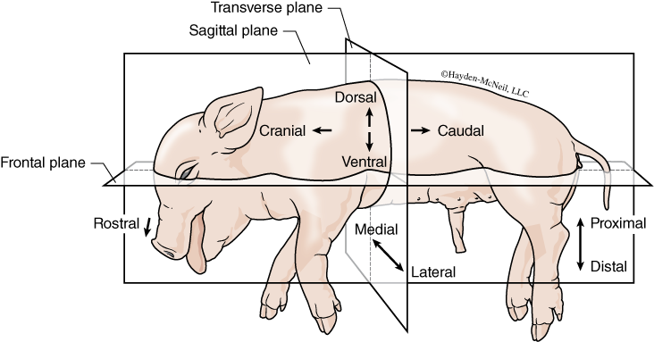
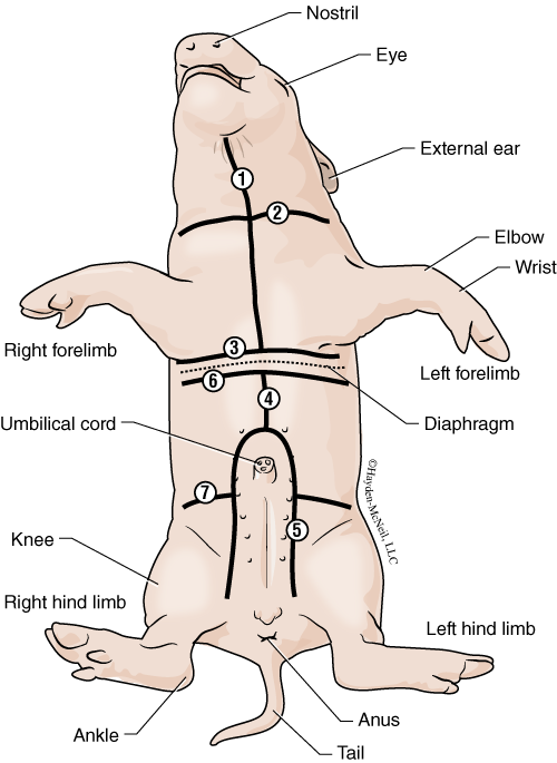
As with all vertebrates and other bilaterally symmetric animals, the fetal pig shows cephalization, or a concentration of the nervous system and sensory organs in the head. In contrast, animals such as a sea anemone, a starfish, or even a clam lack cephalization. In a bilaterally symmetrical animal, the head region is referred to as anterior or cranial (Figs. 3.1 and 3.2). The tail end region is posterior or caudal. Dorsal refers to the back of the animal. In vertebrates, the vertebral column is dorsal. The opposite side is ventral. The term proximal is a relative term that means closer to the median plane of the animal than some other structure. Distal is a relative term that means further away from the middle of the animal. For example, your wrist is distal to your elbow, but the elbow is proximal to your wrist. Lateral is also a comparative term that means away from the middle. Medial means toward the middle, or midline, of the animal. The terms left and right always refers to the pig’s left or right side (not your left or right). Superficial refers to structures closer to the surface; deep refers to structures away from the surface. The standard anatomical position is shown in Fig. 3.7. Humans are an atypical vertebrate because we are bipedal (most mammals are quadrupedal) and for us, the terms anterior and posterior are used interchangeably with ventral and dorsal.
PROCEDURE
For your work with the fetal pig, you will be working in groups of 2 or 3 over the next several weeks. Your TA will give you the pig today and give you instructions on how to label it (your name and section number), and where the pig will be kept.
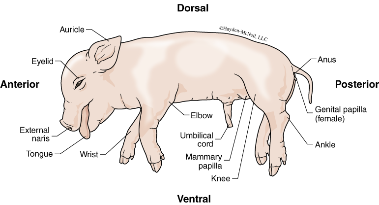
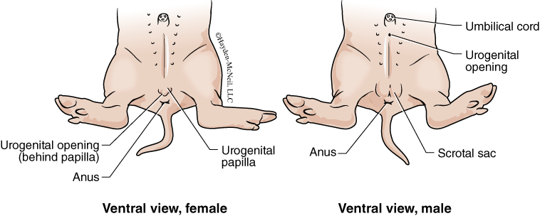
First, familiarize yourself with the external anatomy of the pig. You should be able to find and know all of the structures listed in Fig. 3.2. In addition, you must be able to recognize both male and female pigs (Fig. 3.3) by their external structures. In males, the urogenital opening is just posterior to the umbilical cord. The scrotal sac (which may be more or less conspicuous, depending on its stage of development) is anterior to the anus. In the female, the urogenital opening is anterior to the anus and posterior to the urogenital papilla. Be sure to look around the room at other pigs to see several examples of males and females.
Make a quick sketch of the fetal pig for your notebook and label it with the terms indicated in Figs. 3.2 and 3.3. In addition, you should indicate the dorsal and ventral side, the anterior and posterior end.
Next, you and your partner should quiz each other using the anatomical terms on the previous slide. You can do this by guiding your partner’s probe by verbal cues, using terms such as proximal and distal, ventral and dorsal. By the end of this period you should be comfortable with these terms.
As we go through the various organ systems in the next few weeks, it is very important to keep in mind that they are all interconnected!
III. DIGESTIVE SYSTEM
Not all animals have a digestive system, but all animals are heterotrophs and require food from outside of their bodies. Sponges and tapeworms completely lack a digestive system, while several groups such as the Hydra, anemones, and flatworms have a digestive “system” called a gastrovascular cavity with a single opening. The majority of animals (including arthropods, many worms, and vertebrates) have a digestive system that is sometimes called a “tube within a tube.” That is, the body itself is a tube and the digestive system is also a tube located within the bigger tube (the body). The digestive system has an anterior end (the mouth) and a posterior end (the anus). This tube has specialized regions, and each of these regions has very particular functions all related to digestion. In this lab we will be focusing on this kind of digestive system. We will be examining the mouth, esophagus, stomach, intestines, and anus, as well as organs and glands associated with the digestive system.
The function of a digestive system is to do all that is necessary to get organic material that is outside of the body into the form where it can be used by individual cells inside the body. For animals that are predators, this would include teeth and jaws that allow an animal to kill another. For animals that are herbivores, such as a cow or a horse, this would include molars that are very good at grinding plant material. Other animals are omnivores who can eat both meat and vegetation. The digestive system both mechanically and chemically breaks down food entering the body, and by connections with the circulatory system and the lymphatic system, this material is absorbed and taken to the liver and the rest of the body.
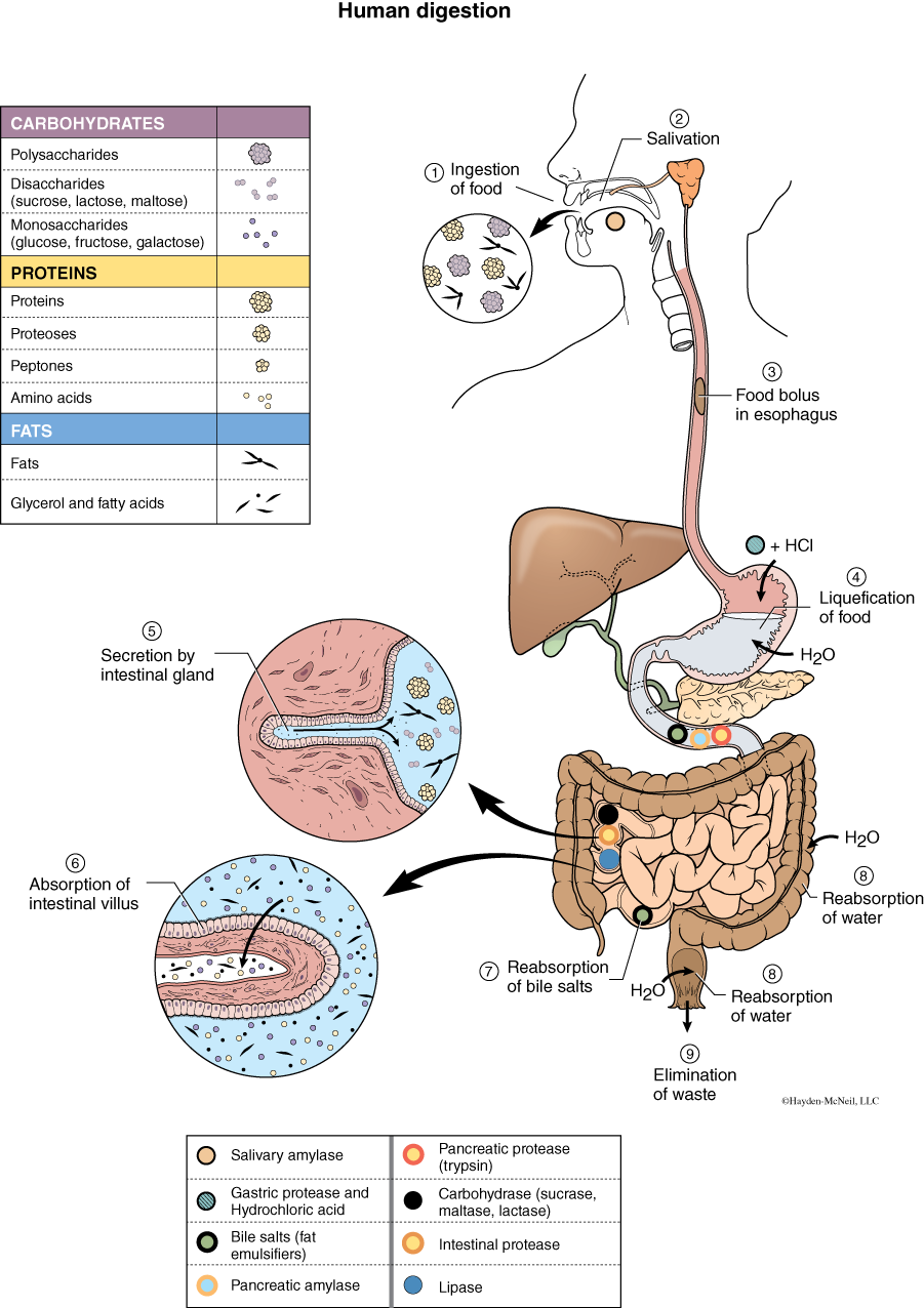
Often, digestion is divided into these processes: mechanical and chemical digestion, secretion, and absorption. From this laboratory, from lecture, and from your reading, you should know where these processes occur. We will start with a brief description of the organs involved in digestion and then provide detailed instructions for the dissection.
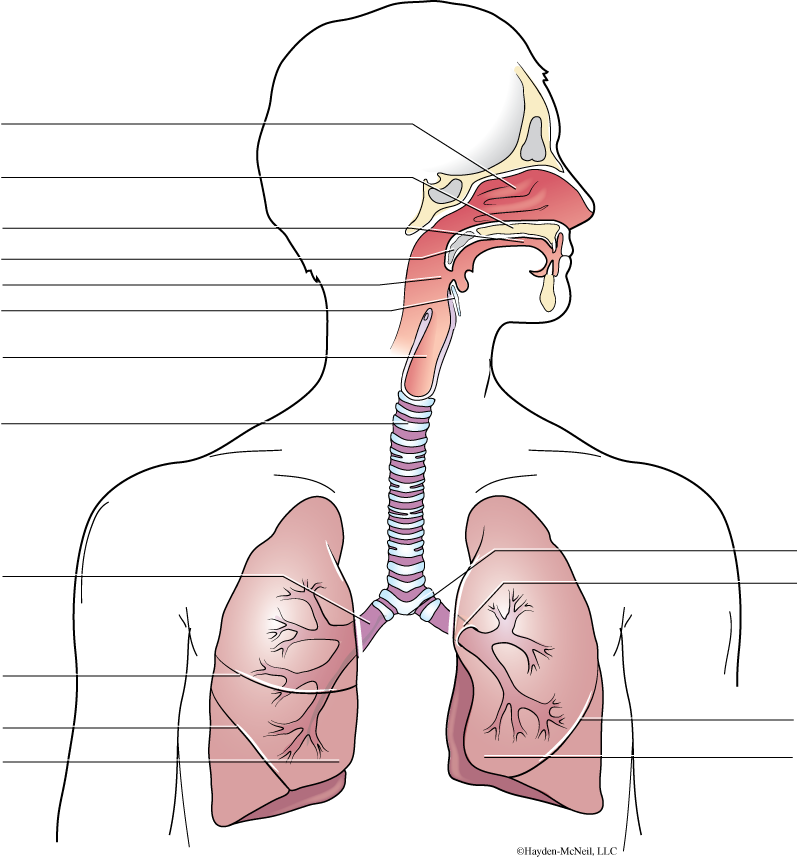
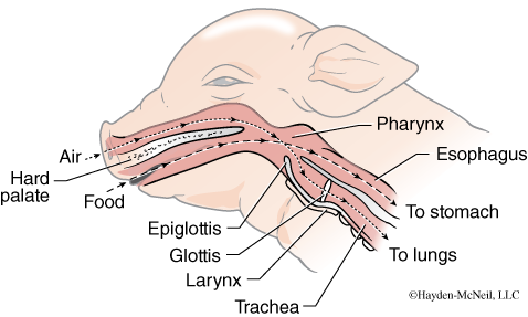
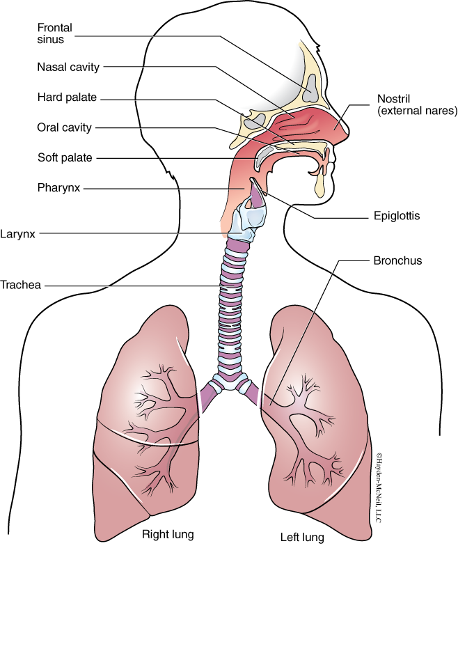
The front end of the digestive system is the mouth. In the mouth, food is mixed with saliva from the salivary glands. The saliva functions to both moisten the food and to start the process of chemical digestion. Thus, in the mouth, both chemical digestion (from the action of enzymes) and mechanical digestion (from chewing) has begun. The tongue is important in tasting and in moving food around the mouth. Once chewed, the food moves to the back of the throat to the pharynx and is swallowed as a moist bolus of food. The pharynx (or nasopharnyx) is a region where both food and air passes, going toward the stomach and the lungs. When swallowing occurs, a flap called the epiglottis closes over the opening to the trachea (cartilaginous tube going to the lungs), and is an important means of keeping food going down the esophagus (muscular tube going to the stomach), and not going down the trachea.
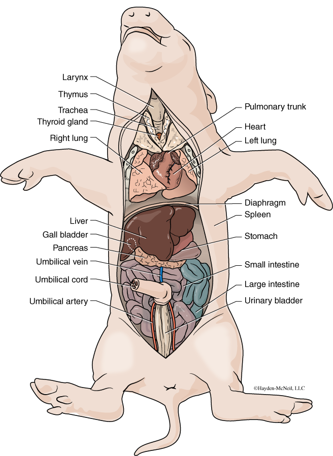
Although not part of the digestive system, we will see the thymus and thyroid today and so their description is included here. In the neck, ventral to the trachea, is the thymus gland (Fig. 3.8). The thymus gland is very large in fetuses and small in mature animals. It is important in the immune system, as it is where the T-cells (a type of white blood cell) mature and become active. The thymus typically covers the anterior portion of the heart and extends anteriorly along the trachea. Beneath the thymus (dorsal to the thymus) is the thyroid gland, a small solid oval mass. The gland secretes thyroxine and is a very important endocrine structure (see Lab 6).
The esophagus is a muscular tube that extends from the pharynx to the stomach. It is an organ with smooth muscle, and once swallowing occurs, the movement of the food is not under conscious control. A peristaltic wave of contraction moves down the esophagus, pushing food into the stomach. The esophagus passes through the diaphragm, a large muscle that separates the thoracic cavity from the abdominal cavity. The stomach lies on the left side of the pig, under the large dark liver. The stomach in many animals is large enough to store food for a period of time, allowing the animal to eat when food is present and digest it shortly thereafter. It also secretes hydrochloric acid and pepsinogen, an enzyme that works well in an acid environment and helps to break down protein. There is a sphincter (muscular ring) on both ends of the stomach controlling material entering and exiting the stomach. The cardiac sphincter connects the esophagus to the stomach, and the pyloric sphincter connects the stomach to the intestine.
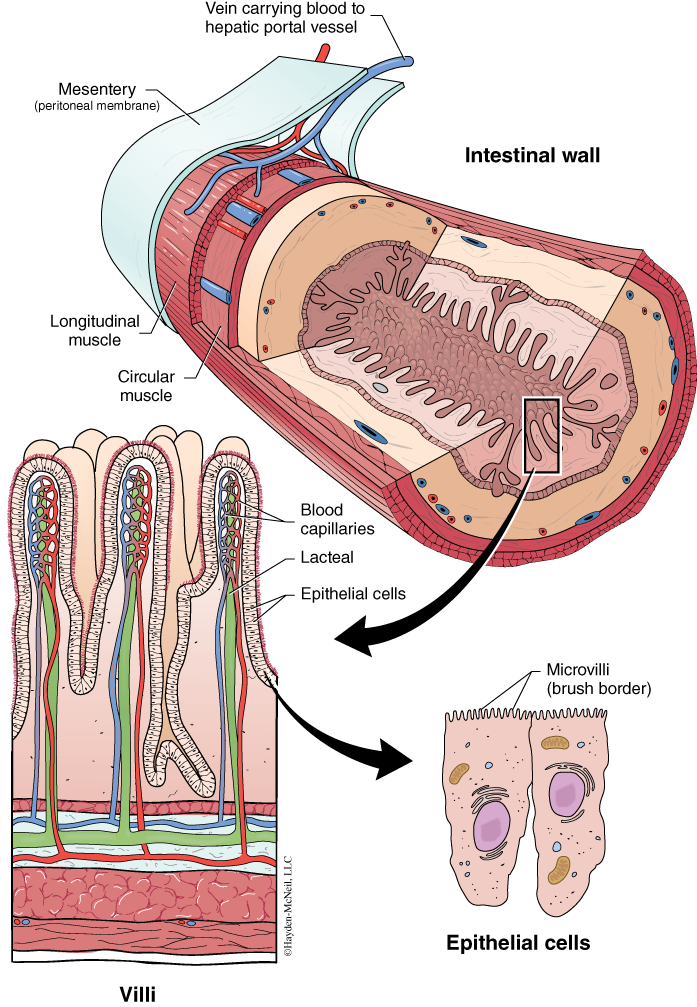
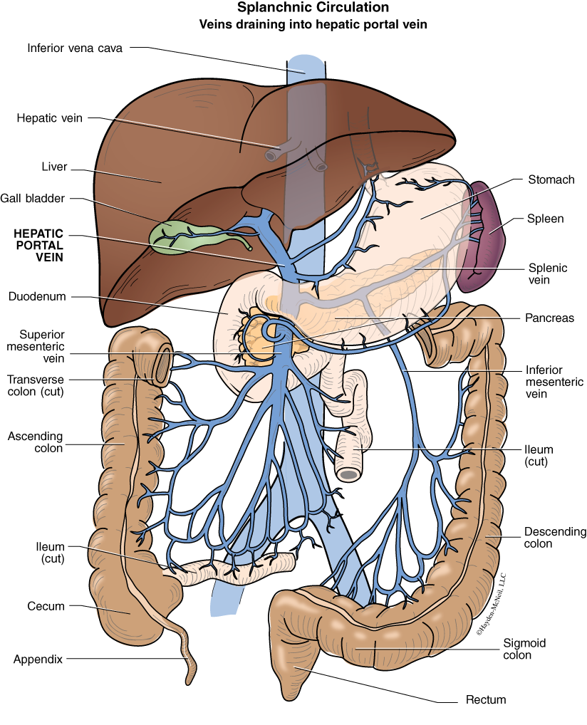
The first portion of the small intestine is called the duodenum. The duodenum and the other sections of the small intestine, the jejunum and ileum, are held by mesenteries. Mesenteries are sheets of connective tissue that hold organs in place and provide a pathway for blood vessels and nerves. The small intestine is where most of the absorption of nutrients occurs. The inside of the small intestine has millions of tiny finger-like extensions, called villi, that extend into the lumen of the intestine. In addition, each villus is comprised of many cells, and these cells have microscopic extensions called microvilli. The villi and microvilli vastly increase the surface area for absorption. In the middle of each villus is a structure called a lacteal (a lymphatic capillary), where fats are absorbed. Mesenteric veins connect with the capillaries that are around the villi and then connect with the hepatic portal vein. Nutrients absorbed from the small intestine are taken to the liver via this system.
Where the small intestine connects to the large intestine, there is a blind pocket of the gut, called the cecum. In many herbivores, the cecum is large and houses both bacteria and protists that are important in digesting cellulose. In humans, this pocket is small and is called the appendix.The large intestine or the colon is responsible for reabsorption of water and the production of feces. It is substantially larger in cross section than the small intestine. Finally, the end of the digestive system is the rectum and the anus. There is another sphincter at the anus.
Immediately beneath the diaphragm is the very large liver, one of the largest organs in the abdomen. The liver has many important functions in digestion, including the production of bile, the conversion of glucose to glycogen and glycogen to glucose, and the detoxification of many compounds from the food. The liver also converts glycerol and fatty acids to compounds used as fuel in cellular respiration. The liver removes excess amino acids from the blood and breaks them down by deamination, forming ammonia and then urea from the ammonia. Urea is a nitrogenous waste that is then removed by the kidneys. Working with the spleen, the liver also is important in the breakdown of old blood cells.
The gall bladder is a small, round, greenish organ on the underside of the right lobe of the liver. It stores bile that is secreted by the liver. Bile enters the duodenum via the cystic duct. Bile salts help digestion by emulsifying fats. The other component of bile is the pigment bilirubin, which is a byproduct of the breakdown of hemoglobin in old blood cells, something that occurs in the spleen and the liver.
The pancreas is a gland that, among other functions, secretes digestive enzymes into the duodenum. It is elongate and glandular and is embedded in the mesenteries that support the stomach. The are two lobes to this gland. Digestive enzymes travel down a small duct that connects to the duodenum. The pancreas is also an endocrine organ that produces the hormones insulin, glucagon, and somatostatin. The former two are important in regulating blood glucose levels; somatostatin is important in regulating insulin and glucagon.
The spleen is another organ found near the stomach. It is long and thin and has a texture similar to the liver. It is a storage organ for blood cells and is instrumental in breaking down old blood cells.
PROCEDURE: SALIVARY GLANDS, MOUTH, AND NECK REGION
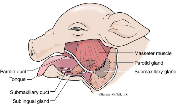
Begin your dissection by examining the salivary glands on the side of the face of your pig. By dissecting and removing the skin and teasing apart the connective tissue beneath, you should be able to find the large, right masseter muscle, the right parotid and submaxillary gland, and possibly the right sublingual gland and parotid duct. These salivary glands are all comprised of epithelial tissue. They secrete the saliva, a fairly complex liquid that starts the process of chemical digestion and also moistens the food for its passage through the animal. In your laboratory notebook, you should make labeled sketches of what you find and how it looks.
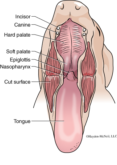
Next you will look at the oral cavity of your pig. Open the mouth wide and, using your scissors or scalpel, cut from the edges of the mouth toward the ear to open the mouth wide. Note (and be careful of!) the incisors and canine teeth. Find all of the structures labeled in Fig. 3.5. Note that the epiglottis covers the trachea when the animal swallows, allowing food to go down the esophagus and not the trachea. When the animal is not swallowing, the epiglottis does not cover the trachea, allowing for normal breathing. Fig. 3.6 shows a lateral view that clarifies the relationship between the pharynx, esophagus, and trachea. Use a blunt probe to explore the nasopharynx region and to see both the esophagus and trachea.
PROCEDURE: GETTING INTO THE BODY CAVITY
The next step is to get into the body cavity of the pig. Fig. 3.7 shows the order and position of cuts that allows one to get into the body cavity. This is best done with scissors and it works well to start near the chin and work toward the intersection of lines 1 and 3. You will cut through skin and muscle, but try not to go deeper at first. Next make the lateral cuts #2 and #3.
Be very careful in your cut around the umbilical cord! Cut #4 starts just anterior to the umbilical cord, and as you cut cranially, you should find the diaphragm. Cut #5 must be a careful cut around the umbilical cord. At this point you want to keep all of the connections to the umbilical cord intact. Cuts #6 and #7 help to open up the abdominal cavity. You may see excess latex from where vessels burst. (This is more often veins than arteries. Why?) If there is excess fluid in the cavity, it can be put in the sink. If there is excess latex, it should be gently removed and thrown away. If there is coagulated blood, it can be gently rinsed and removed.
In the neck region you should first find the thymus gland (Fig. 3.8). The thymus covers the anterior portion of the heart and extends anteriorly along the trachea. Beneath the thymus (dorsal to the thymus) is the thyroid gland, a small, solid, oval mass. In the neck, find the trachea and the esophagus. How do they differ? Where is each located? Make an incision in the trachea and insert your blunt probe to see where it comes out in the throat. Likewise, make an incision in the esophagus and trace that to the mouth and to the stomach.
You should next find the diaphragm, a large muscle that separates the thoracic cavity from the abdominal cavity. The esophagus passes through the diaphragm, connecting the mouth with the stomach. Immediately beneath the diaphragm is the very large and prominent liver. The stomach lies on the left side of the pig, under the liver. Find the cardiac sphincter (connecting esophagus to the stomach) and the pyloric sphincter (connecting the stomach with the small intestine).
Cut open the stomach and look for the folds called rugae. With your blunt probe examine the cardiac sphincter and the pyloric sphincter.
The first portion of the small intestine is called the duodenum. Find the duodenum and the mesenteries that hold it in place. Find the mesenteric veins and the hepatic portal vein that transport blood from the capillaries around the intestine to the capillaries in the liver.
Find and sketch the gall bladder, a small, greenish, round organ on the underside of the right lobe of the liver. It stores bile that is secreted by the liver. Find and sketch the spleen; it is another organ found near the stomach. It is long and straplike in the fetal pig and has a texture similar to the liver. What is the function of the spleen?
The pancreas is a gland that, among other functions, secretes digestive enzymes into the duodenum. It is elongate and glandular and is embedded in the mesenteries that support the stomach. There are two lobes to this gland. Digestive enzymes travel down a small duct that connects to the duodenum (this can be difficult to find).
Return to the small intestine, cut a small opening in the intestine, and look at the inside texture. What does it look like? Snip a small piece, put it in a dish of water, and look at it with a dissecting scope. Sketch what you see. Locate the villi. Next, get a prepared microscope slide and examine the details of the cross section of the intestine. Sketch and label your drawing. Compare your drawing with Fig. 3.9. The three regions of the small intestine are the duodenum, the jejunum, and the ileum.
Next find the cecum, and the place where the small intestine joins the large intestine. It is substantially larger in cross section than the small intestine. You should examine the inside texture of the colon and look at it with the dissecting scope. How is it similar to the small intestine? How is it different? Finally, find the anus.
Although there are some differences in shape and location, the same organs are present in humans. A summary of the organs and the processes in humans is shown in Fig. 3.10. Fig. 3.11 shows in humans how the hepatic portal vein connects the capillaries surrounding the intestines with the capillaries in the liver. By definition, a portal vein joins two capillary beds.
IV. RESPIRATORY SYSTEM
Not all animals have a respiratory system, but all animals do need to get oxygen to all of their cells. How is this accomplished? Small and very small animals (rotifers, nematodes, flatworms) can rely on diffusion of oxygen and carbon dioxide directly across their cuticles. Larger animals require structures that help them to increase the surface area for diffusion. Some aquatic animals have gills, as is seen in many fish and also some insects. Terrestrial animals have a variety of structures including lungs and trachea, as is seen in mammals and birds, or just trachea, as is seen in terrestrial insects, or a combination of structures such as book lungs and trachea, as is seen in many spiders. In all of these cases, the primary function of the system is to provide surface area for diffusion. In many animals (but not insects or spiders) the respiratory surface is closely associated with the circulatory system.
In mammals, the organization of the respiratory system is similar across species, including humans and fetal pigs (Figs. 3.8 and 3.12). In this lab, we will look at the not yet functional respiratory system of the fetal pig and compare it to the system in humans.
The respiratory system of mammals opens to the outside at the mouth and nose. In the fetal pig, air enters primarily through the nostrils. The passage of air across moist membranes in the nose helps to moisten the air and to filter large particles from the air. The epithelial cells that line the nasal passage secrete mucus, a very sticky substance that helps to trap particles and bacteria. These cells are ciliated, and the cilia help to move the mucus to the back of the throat where it is swallowed and taken to the stomach.
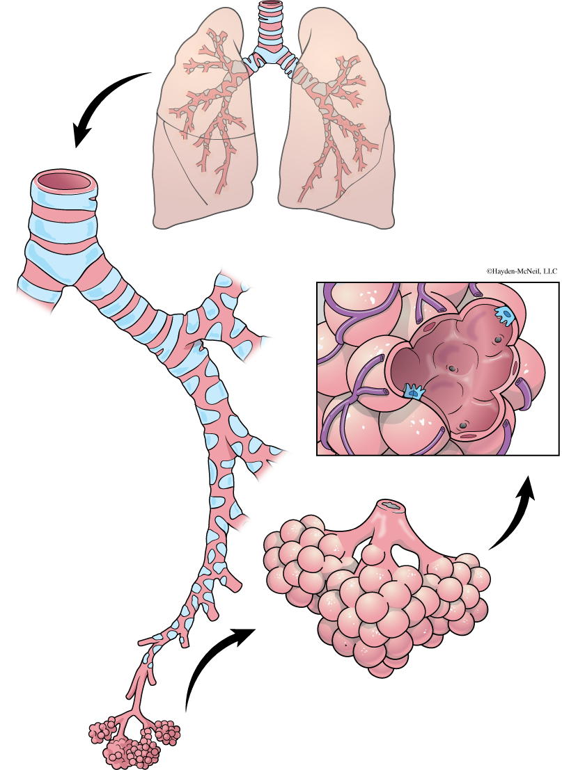
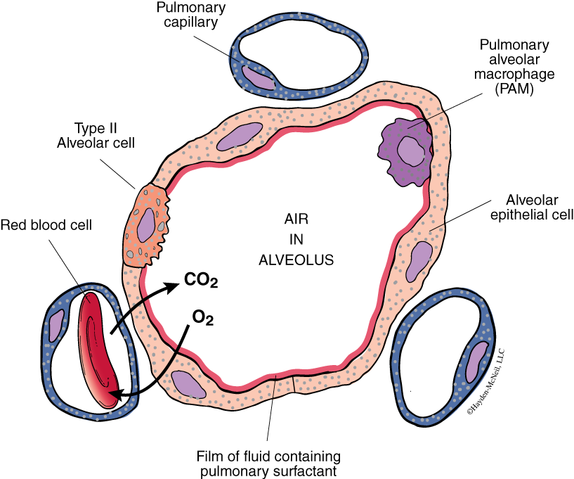
The trachea is a tube that is reinforced with cartilage that runs from the nasopharynx to the lung. It is ventral to the esophagus and because of the cartilage, it is easier to find and trace. The trachea leads through the larynx, a structure that allows mammals to make a wide variety of sounds, including speech in humans. When it gets close to the lungs, the trachea splits into two bronchi—a right bronchus and a left bronchus—which lead to the lungs. The right lung has four lobes, while the left lung has three. Inside the lungs, the bronchi subdivide again to bronchioles. The bronchioles also subdivide into smaller and smaller tubes, until each part terminates in a microscopic sac called an alveolus (see Figs. 3.13 and 3.14). Each alveolus is encircled with capillaries, and it is here there is the exchange of oxygen and carbon dioxide.
PROCEDURE
Earlier in this lab you dissected the mouth and pharynx of the fetal pig. You should review that now to be clear about how the esophagus and trachea join at the pharynx. You will follow the trachea as it goes to the lungs. The trachea is a tube that is reinforced with cartilage. It is ventral to the esophagus and because of the cartilage, it is easier to find and trace. Find the larynx and follow the trachea to where it divides to the two bronchi. Find the four lobes of the right lung and three lobes of the left lung. Follow a single bronchiole as far as you are able.
Cut a small piece of lung, place it in a small dish with water, and look at it with the dissecting scope. Sketch what you see.
Finally, examine and sketch a prepared cross section of a lung, showing the alveoli.
How does the fetal lung differ from an adult lung?
V. NOTES TO INSTRUCTORS AND MATERIALS FOR LAB
This lab starts a several-week sequence where we will be dissecting the fetal pigs. It is important to provide the framework for students to succeed at this important task. Students must bring their own dissecting kit, gloves, and goggles to each of these labs.
Be sure to give clear instructions about dissections.
Take time during the middle or near the end of the period for students to look at other pigs. This both gives them the opportunity to explain their dissection to other students and to see other examples. This needs to be a regular event for all dissection labs! You can stop everyoneand say, “now is the time to compare with other pigs.”
EXTERNAL ANATOMY
Know anatomical planes of reference and regions of the body. Know all labeled features of the fetal pig. Be able to distinguish sex in the fetal pig.
DIGESTIVE SYSTEM AND ORGANS
Students should dissect the salivary glands and the oral cavity, as well as the thoracic and abdominal cavities. Know the function of all organs. Students should create a table listing all organs in the digestive system and their functions.
RESPIRATORY SYSTEM
Know the function of all organs and know and understand how they are connected.
Complete the attached worksheet.