Chapter 4. CIRCULATORY SYSTEM
I. OVERVIEW
The circulatory system is the major connecting system within the vertebrate body. How does food get from the digestive system to the liver? The circulatory system. How does oxygen get from the lungs to the big toe? The circulatory system. How do hormones move from where they are formed and secreted to their final destination? You got it…the circulatory system. The circulatory system takes nitrogenous wastes to the kidneys and is of key importance in water regulation. In mammals (and other vertebrates), the circulatory system has many functions: transport of gases such as oxygen and carbon dioxide, transport of nutrients and metabolic wastes, transport of water and other materials to the interstitial fluid, and transport of hormones. In many animals, the circulatory system interacts with every other organ system. It is also a critical component of the immune system and is critical in homoestatic mechanisms, such as maintaining a correct temperature.
However, not all animals have a circulatory system. For example, Hydra, corals, and flatworms completely lack a circulatory system. Typically, it is smaller animals (like a Hydra) that completely lack a circulatory system. Insects and spiders and many molluscs have a circulatory system, but it is fundamentally different from that seen in vertebrates. Unlike the vertebrates that have a closed circulatory system (a system where the blood never leaves the vessels), the insects, spiders, crustacea, and many molluscs have an open circulatory system: they have a heart and arteries, but no capillaries or veins. At this point in the semester, we will focus mostly on the vertebrate system, using the fetal pig as our model.
As mentioned above, vertebrates have a closed circulatory system, a system made up of a series of tubes (arteries and veins) and a fluid (the blood). In this lab we will focus on both the “plumbing” (the vessels) and on the blood. The vessels of the system are comprised of a pump (the heart), vessels that carry blood away from the heart (arteries), smaller vessels connected to the arteries (arterioles), and smaller vessels still (capillaries). Capillaries are microscopic and in many, the blood cells pass through them one at a time. The capillaries are connected to small vessels (venules) that carry blood back toward the heart and that join with the larger vessels known as veins. We will examine the fundamental differences between arteries and veins.
In this lab, we will start with a dissection of a sheep heart. It is similar in size to a human heart and is big enough to see. We will learn the path of the flow of blood through the heart. We will then view the fetal heart and compare the circulation through the fetal heart with the circulation through the adult heart. Next, we will dissect anterior and posterior vessels, and we will look at blood cells and at cross sections of arteries and veins. Finally, we will review the specifics of how the circulatory system intersects with other organ systems.
II. THE SHEEP HEART (ADULT CIRCULATION)
The heart is an amazing muscle. In the course of this lab period, on the average, a human heart will beat 12,600 times! In the course of a day it will beat 100,800 times; in the course of a year (five hundred twenty-five thousand six hundred minutes), an average human heart is likely to beat nearly 37 million times.
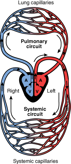
To understand the flow of blood through the heart, it is helpful to see that in mammals there are two separate circuits of blood (Fig. 4.1). One is called the pulmonary circuit, in which de-oxygenated blood travels from the heart to the lungs and back to the heart; the second is called the systemic circuit, where blood is pumped from the heart to the rest of the body and back to the heart.The heart has a right and left side, and each side has an atrium and a ventricle. Thus, there are four chambers in a vertebrate heart. The right side of the heart is integral with systemic circulation. Valves between the chambers assure that blood flow is one direction through the chambers.
To understand the pulmonary circuit, we will start with the right atrium. The right atrium receives deoxygenated blood from the body. Blood enters the right atrium, passes through the tricuspid valve, and goes to the right ventricle. From the right ventricle, deoxygenated blood is pumped to the pulmonary trunk, which branches into the right and left pulmonary arteries, and is then taken to the capillaries of the lungs. The blood returns oxygenated to the left atrium via the pulmonary veins.
The systemic circuit starts with the left atrium. From the left atrium, blood flows to the left ventricle, the largest of the chambers. From the left ventricle, blood is pumped out the aorta, through the aortic arch and to the rest of the body, distributing nutrients and picking up wastes. After passing through capillary beds throughout the body, veins return blood to the right atrium, and we are again ready to go through the pulmonary circuit.
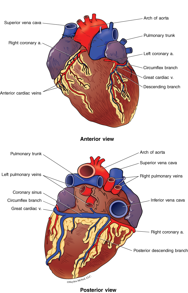
Since the heart is muscle and since it is working all of the time, it must be supplied with blood as well. Fig. 4.2 shows an external view of the anterior side and posterior side of the heart. The coronary arteries and coronary veins that provide oxygen to the heart are visible on both sides of the heart. Damage to these vessels limits the oxygen getting to the heart muscle. If oxygen is deprived for more than a few minutes, damage to the heart muscle occurs.
4.1 PROCEDURE
For this next procedure we will work in groups of two or three. Each group will get a sheep heart. Start by getting oriented as to anterior and posterior, left side and right side. Recall that the terms left and right refer to the heart’s left and right. Next find the right and left coronary arteries on the anterior surface of the heart. Find the pulmonary trunk (Fig 4.2), which should be a prominent vessel on the anterior, cranial end of the heart. The pulmonary trunk will divide into the right and left pulmonary arteries. Often, these vessels (and others) are difficult to identify because they are very close to the heart.
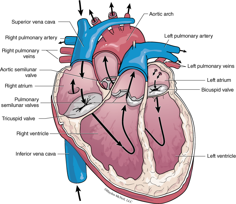
Next we will look inside the heart. Your TA will show you where to make the cut to best see all the chambers. And, after the first lab does this dissection, we will not need to do any more cutting. Find the right ventricle, which is connected to the pulmonary trunk (Figs 4.2 and 4.3).
Find the right ventricle and pulmonary artery. Note that when the right ventricle contracts, blood is forced out of the pulmonary artery, but prevented from going backwards to the right atrium by the tricuspid valve (Fig. 4.3). When leaving the right ventricle, blood passes through the pulmonary semilunar valve and enters the pulmonary artery, which divides into the right pulmonary artery.
Use your blunt probe to trace the movement of the blood from the right ventricle to the pulmonary artery.
Blood returns to the heart from the lungs via the right and left pulmonary veins. Blood enters the left atrium of the heart and passes through the bicuspid valve to the left ventricle. When the left ventricle contracts, blood is forced through the aortic semilunar valve and into the aorta.
Compare the thickness of the wall of the left ventricle with the right ventricle. Which is thicker, and why?
When blood passes through the aortic arch, it is carried both anteriorly and posteriorly, literally to all of the capillary beds in the body. When it returns to the heart, it does so through the venules, which connect to form veins. The anterior vena cava carries blood from the anterior of the body to the heart; the posterior vena cava returns from the posterior of the body to the heart. In humans, because of our upright stature, the anterior vena cava is called the superior vena cava and the posterior vena cava is called the inferior vena cava.
The anterior and posterier vena cavae enter the heart at the right atrium. From the right atrium the blood is forced through the tricuspid valve to the right ventricle and then back to the lungs.
You should use the blunt probe to trace the flow of blood through the heart. When you can accurately show your partner the flow of blood, raise your hand to demonstrate to your TA that you know the flow and can correctly identify all chambers, valves, arteries, and veins.
Last but certainly not least, examine the much larger cow heart on the side table.
III. FETAL HEART AND UMBILICAL CORD (FETAL CIRCULATION)
We will now look at the circulation within a fetus and compare the fetal circulation with the adult circulation. We will discuss both humans and fetal pigs.
The placenta is an organ made of a combination of maternal tissue and some fetal tissues through which gases and nutrients are passed from mother to fetus. The placenta also secretes hormones. Normally, there is not an exchange of blood cells between the mother and the fetus. However, a wide range of materials in solution (including drugs) can pass between the mother and the fetus. The placenta is a mass of capillaries.
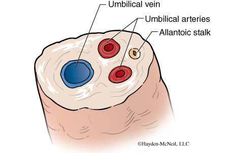
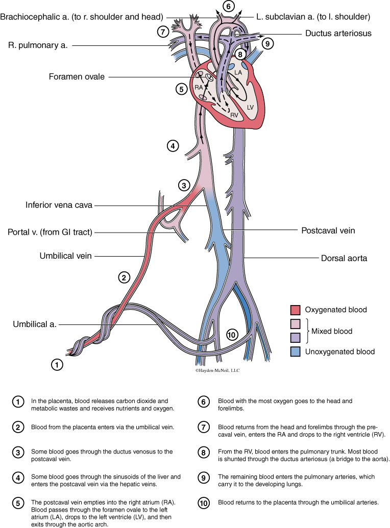
The umbilical cord grows early in the fetus and connects the fetus to the placenta. Within the umbilical cord are two arteries that carry deoxygenated blood away from the fetus, and one vein that carries oxygenated blood back to the fetus from the placenta (Figs. 4.4 and 4.5). The allantoic stalk within the umbilical cord connects to the allantois, a sac that acts as a dump for metabolic wastes of the fetus.
The umbilical vein comes from the placenta and joins the posterior or inferior vena cava at the ductus venosus (which passes under the liver), and brings blood to the right atrium and then to the right ventricle. When the fetal heart contracts, blood is directed from the right ventricle to the pulmonary artery. So far, this is very similar to adult circulation. However, a shunt or connection exists between the pulmonary artery and the aorta, called the ductus arteriosus, so that much of the blood that is oxygenated by the mother passes to the aorta and thus to the rest of the body. A small amount goes to the lungs, and is enough for the lungs to continue to develop. Recall that in the fetus, the lungs are not filled with air. In addition, there is an opening in the septum between the right and left atria called the foramen ovale. This allows some of the blood in the right atria to go directly to the left atria and not go to the lungs. This opening generally closes completely within the first year after birth. However, in about 30% of adult humans there remains a small opening. According to some sources, these people are more likely to suffer from migraines.
PROCEDURE
Examine the umbilical cord of the fetal pig and find and sketch the umbilical arteries and umbilical vein. Which vessels carry oxygenated blood? Which carry deoxygenated blood? Trace the flow of blood from the placenta through the body and back to the placenta. You should write out the major vessels involved in this circulation. Find the ductus venosus.
Next, if you have not already done so, carefully expose the heart on your fetal pig. You may need to cut through the pericardial membrane that surrounds the heart. Be as careful as you can be with all of the vessels associated with the heart. Work to expose the structures without damaging the vessels. Find and identify the right and left ventricle, and the right and left atria. Find the aortic arch as well as the cranial or anterior vena cava. Typically in the fetal pig, the arteries will be whiter and stiffer than the veins, which are thin and often collapsed. Also, veins are more frequently damaged during the process of injecting the pigs with latex. (Why would that be?) A ventral view of the heart (such as when you first expose the heart) should show the coronary artery. Find the pulmonary artery coming out of the top of the right ventricle.
Next, look for the ductus arteriosus, the connection between the pulmonary artery and the aorta. The ductus arteriosus allows much of the blood from the right ventricle to bypass the lung and join the blood in the left ventricle on its route to the rest of the body. You should be able to describe and explain the differences between fetal circulation and adult circulation.
IV. VERTEBRATE BLOOD VESSELS, ANTERIOR, ABDOMINAL CAVITY, POSTERIOR
In this next section we will focus on finding the major blood vessels in the rest of the body. This shows the connections between different organs of the body. In this section we will combine the description of the vessels along with the dissection instructions.
PROCEDURE
We will start with the arteries and will carefully follow the path of the major arteries. In your dissection, carefully remove tissue that is blocking your view of the vessel, but be careful to not cut through veins. Strive for a clear dissection.
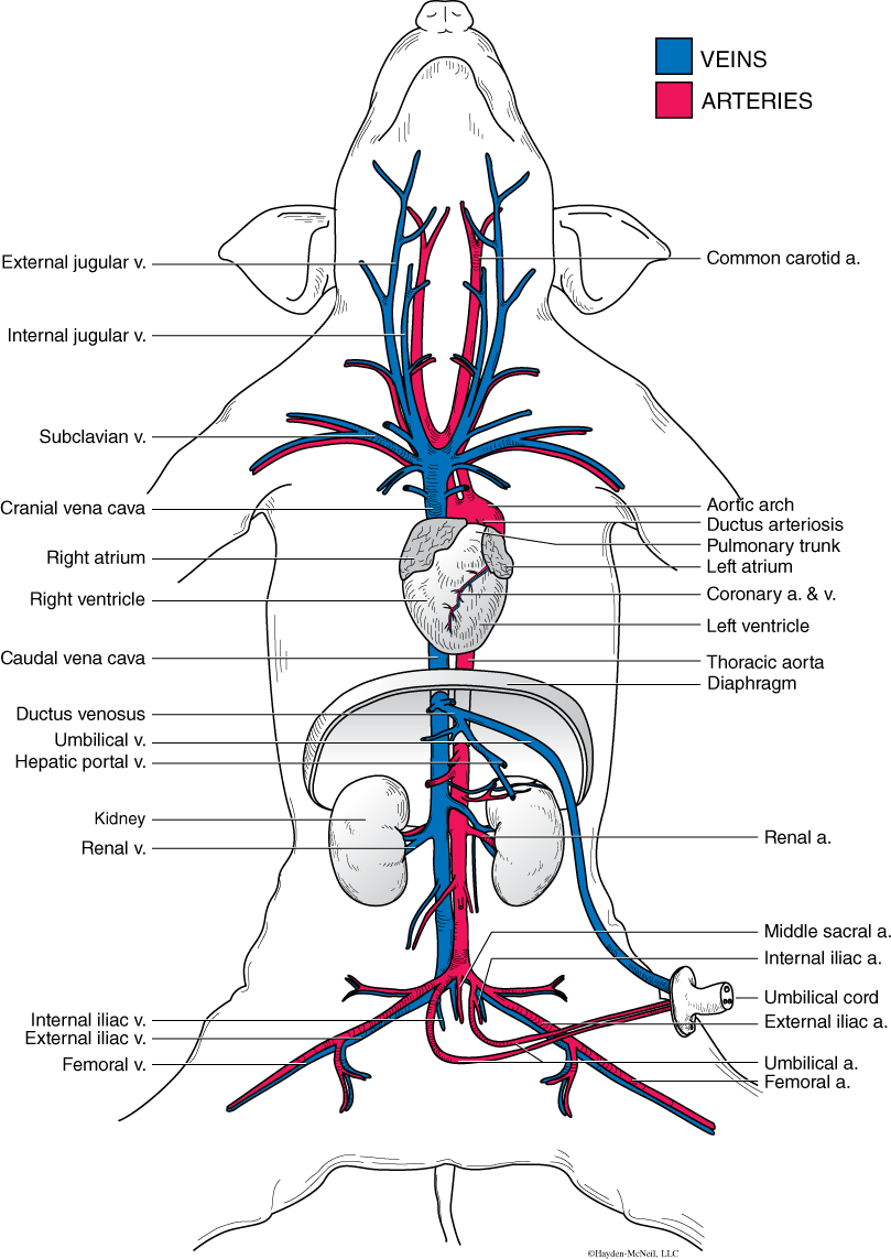
We will start with the veins and arteries of the thoracic cavity (Fig. 4.6). First find the anterior and posterior vena cava and recall that they join at the heart. The anterior vena cava brings blood from the region anterior to the heart, while the posterior vena cava brings blood from the region posterior to the heart. Trace the anterior vena cava anterior (cranially) to its first branch. This very short branch is called the right and left brachiocephalic vein. Continue to trace the veins on one side of the pig and find the four veins that come together to form the brachiocephalic vein. These four veins are the internal jugular, external jugular, cephalic vein, and right subclavian vein. We will provide additional pictures in lab to help you find these veins. Follow the internal and external jugular vein as far as you can.
Earlier you found the large aortic arch coming off the cranial end of the heart. Trace the aorta as it curves caudally. Several branches come off the aorta. The first is the brachiocephalic trunk. The brachiocephalic trunk quickly splits into the right subclavian artery and the common carotid artery. Follow the common carotid artery cranially as far as possible. A second branch off of the aorta is the left subclavian artery.
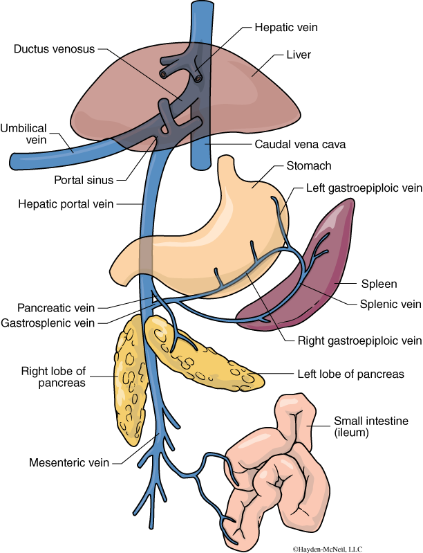
We will next work caudally. Follow the posterior vena cava from the heart to the liver (it will pass through the diaphragm). Under the liver, connecting to the intestines, pancreas, spleen, and stomach, is the hepatic portal system. A portal system is a vein that connects two capillary beds. The hepatic portal system connects the capillary system associated with the digestive organs with the capillary bed of the liver. Materials taken into the blood from the digestive system are therefore taken directly to the liver. Large amounts of sugars and possibly toxins from the digestive system can be filtered by the liver. Recall that the liver can detoxify toxins and also converts glucose to the storage molecule, glycogen. See Fig. 4.7 as a diagram of the human hepatic portal system. Continue to trace the caudal vena cava and find the renal vein and artery.
Trace the aorta caudally, after it arches. Find the celiac artery that goes to the stomach, pancreas, and spleen. Find the renal arteries that go to the kidneys. We will look at some of the veins and arteries in the pelvic region when we examine the reproductive structures.
V. VERTEBRATE VESSELS AND BLOOD
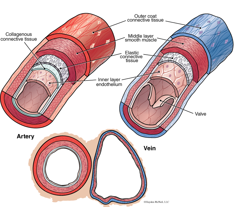
Arteries carry blood away from the heart, and in vertebrates, arteries are muscularized and will contract to help move the blood along. The wall of an artery has three layers: the innermost endothelium (an example of squamous epithelium); the middle layer, composed of smooth muscle and connective tissue; and the outer layer of connective tissue has elastic collagen fibers. Why is it important that the outer layer be elastic? Veins can be recognized by their much thinner walls (Fig. 4.8). Know two examples where arteries carry nonoxygenated blood.
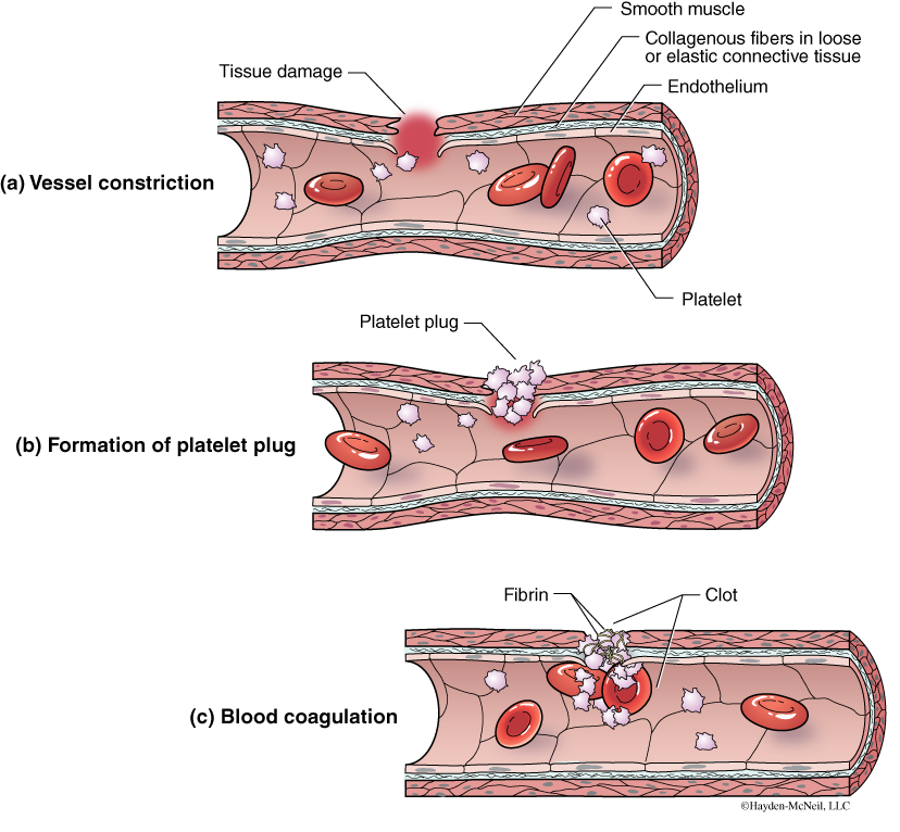
Blood is an example of a connective tissue, as it is made of both cells (red blood cells and white blood cells), a fluid called plasma, and pieces of cells called platelets. Plasma is made of water, salts, nutrients, and a wide variety of other materials that are transported, including plasma proteins. One kind of plasma protein is fibrinogen, a protein important in the clotting process. Platelets are also important in the formation of blood clots (Fig. 4.9).
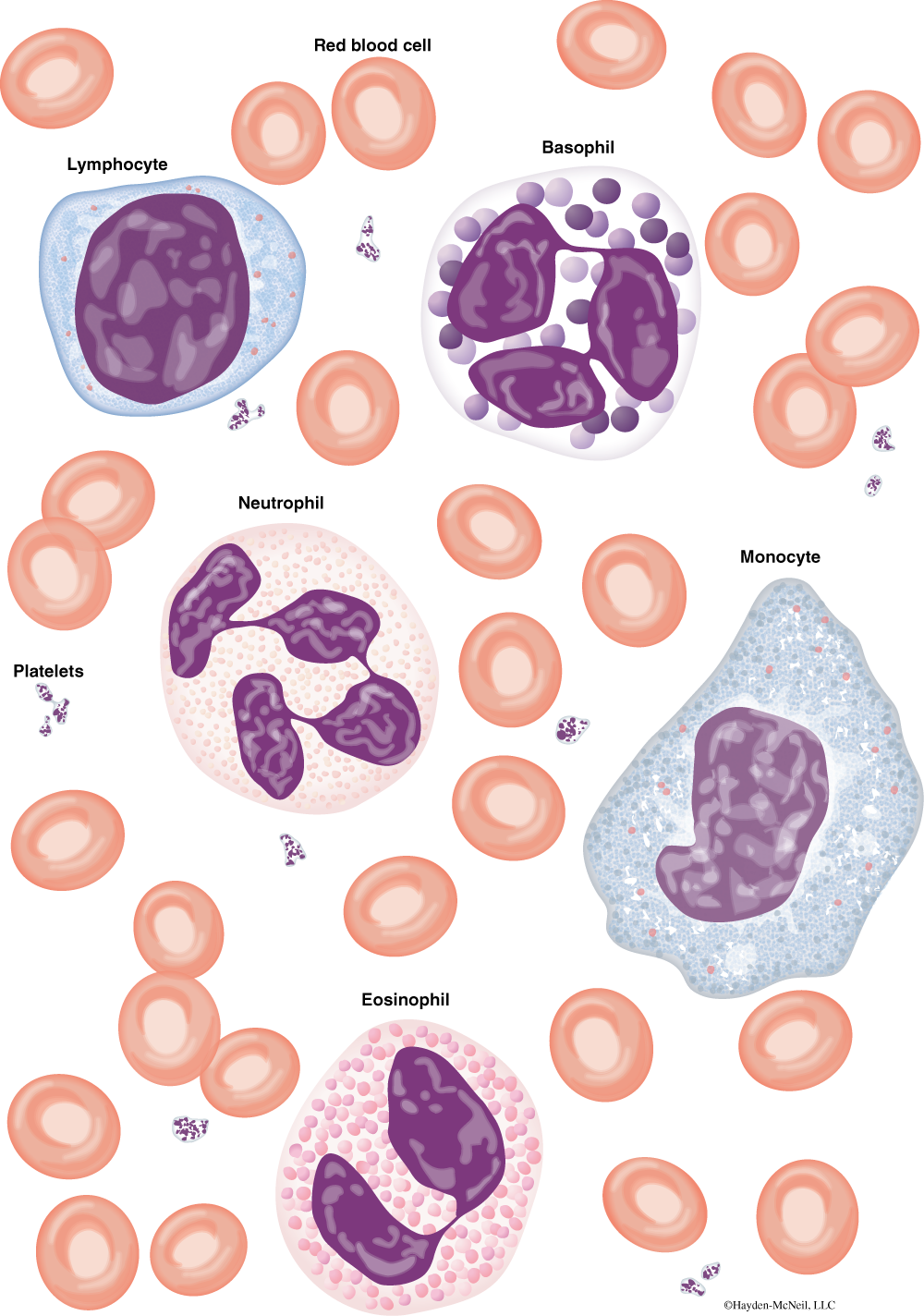
There are two types of blood cells: erythrocytes, or red blood cells, and leucocytes, or white blood cells (Fig. 4.10). The erythrocytes are packed with hemoglobin and are key in the transport of oxygen. The leucocytes are important in the immune response and are specialized in the defense of the body. The leucocytes are divided into two main groups, with several kinds in each group. The granular leucocytes have large, lobed nuclei and granules in the cytoplasm. Examples of granular leucocytes are neutrophils (phagocytic cells that engulf bacteria or dead cells), eosinophils, which seem to be important during allergic reactions and parasitic infections, and basophils, which have granules that contain histamine, a substance that when released causes dilation of the blood vessels. The granules in these leucocytes contain enzymes. The agranular leucocytes are lymphocytes, which produce antibodies, and monocytes, which become macrophages, or giant scavenging cells.
PROCEDURE
From the side table, get a slide that shows the cross section of both an artery and a vein. Make a labeled sketch of each. Be able to recognize each and explain how and why they differ. Label the different kinds of tissue seen in each vessel.
From the side table, get two kinds of blood cells: human blood and frog blood. Examine the human blood first, starting at low power. Move to a high power and look for the different kinds of white blood cells. Do this part of the lab individually, using your own microscope.
It is important that everyone have a microscope and look for these; it is also important that everyone examine at least two different slides.
In your lab book, sketch and identify the different kinds of cells that you can find in the next 5 minutes.
Time yourself for 2 minutes and record how many different kinds of cells you can find in that time:
Eosinophils
Basophils
Monocytes
Lymphocytes
Next, compare the red blood cells of the frog with those of the human. How do they differ in appearance?
VI. NOTES TO INSTRUCTORS AND MATERIALS FOR LAB
This is a full and busy lab. It is also very interesting and fun. Because this is such a full lab, there should be no presentations on this day.
It is helpful to have everyone in the class working on the same thing at the same time. For that reason, you should start with the sheep heart and adult circulation. After 30–45 minutes on this, you should briefly introduce fetal circulation and have students look at the umbilical cord, fetal circulation, and the fetal heart. Next, continue with the dissection, leaving time at the end to look at vessels and blood.
Dissection of the arteries and veins takes time to do carefully.