Chapter 5. REPRODUCTIVE AND EXCRETORY SYSTEMS
I. OVERVIEW
The reproductive system is in some ways one of the most specialized systems in the body. In animals, it is in the gonads where meiosis occurs and haploid gametes are produced. In the vast majority of animals, most of the body is diploid, and only the gametes are haploid. When fertilization occurs, and the gametes join, there is a new combination of genes and the next diploid generation starts to develop. Later in this class, we will contrast the animal model of reproduction with a plant model where there is alternation of generations; one whole portion of the life cycle of the plant is haploid, and another portion is diploid. In plants, gametes are produced by mitosis while in animals, gametes are produced by meiosis.
Sexual reproduction is nearly ubiquitous in animals. However, some kinds of animals can reproduce asexually as well as sexually. And, while in mammals the sexes are separate, in other animals, a wide variety are hermaphroditic (both sexes are present in one individual, as is seen in earthworms), or are sequentially hermaphroditic (as is seen in many fish) and can change their sex depending on circumstances. Since sexual reproduction is so common, there must be a tremendous benefit, as asexual reproduction is more efficient. Most biologists agree that sexual reproduction increases genetic variety in a population and, over time, this increases the fitness of that population.
Some animals, including amphibians and many marine invertebrates, may have external fertilization where gametes are shed directly into the water. Mammals, birds, and reptiles have internal fertilization.
In the Biology Department, Dr. Rich Buchholz studies reproductive behavior in turkeys.His research has focused on female choice of mates and how a male’s condition affects the female’s choice of mates. In addition, he is studying the effects of parasitism on the process of signaling between the sexes. Dr. Stratton studies reproductive behavior in spiders.
In this lab, we will first look at the reproductive structures of the male and female fetal pig. We will look at the details of spermatogenesis and oogenesis and, finally, we will examine the excretory system in the fetal pig.
II. FETAL PIG: STRUCTURES VISIBLE EXTERNALLY
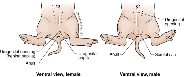
As discussed in the first lab on the dissection of the fetal pig, the males and females are superficially similar. However, with close examination you can see the differences (Fig. 5.1). In the male, the urogenital opening is anterior, near the umbilical cord. In females, the urogenital opening is between the urogenital papilla and the anus. In both, the urogenital opening serves as the opening for both the urinary system and the reproductive system. In males, the scrotal sac, or the sac that holds the testis, should be visible. However, depending on how mature the fetus is, the testes may not have descended into the scrotal sac. In females, the urogenital papilla is homologous with the penis.
III. MALE REPRODUCTIVE SYSTEM AND SPERMATOGENESIS
The main functions of the reproductive system are to produce gametes and to facilitate the joining of the gametes in fertilization. The organs that produce the gametes are the gonads. The male gonads that produce sperm are the testes (sing. testis); the female gonads that produce the eggs are the ovaries. Both the ovaries and testes also produce hormones that are important in reproduction.
In the fetal pig, the male testis is a small, bean-shaped organ. Within the testis is a microscopic coiled tube called the seminiferous tubule, which is where sperm are produced through the process called spermatogenesis. The seminiferous tubule is long and coiled, and in looking at the cross section of a testis, you see a series of circles. The circles are the tubules in cross section (Fig. 5.2).
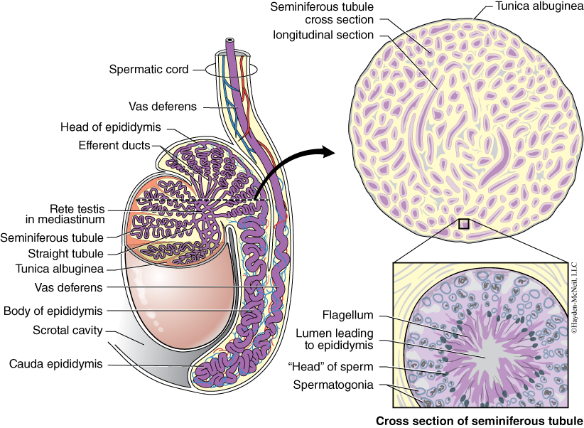
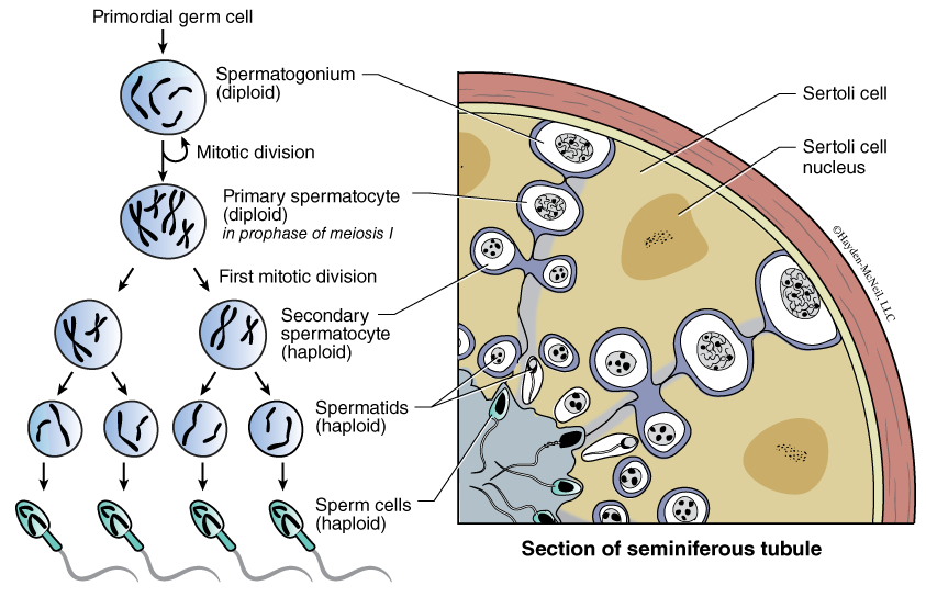
Spermatogenesis is the process of meiosis combined with specific changes that happen in the production and maturation of sperm (Fig. 5.2). The seminiferous tubule provides a large surface area for the production of sperm. Also, within the testis there are specialized cells called interstitial cells that produce testosterone. This is the primary site for testosterone production; however, it is also produced in the ovaries and the adrenal glands. The stages of spermatogenesis are spermatogonium (found near the outside edge of each tubule), primary spermatocyte, secondary spermatocyte, spermatid, and sperm cells (located near the center of the tubule). The cells complete the process of meiosis (and spermatogenesis) as they move from near the outside edge of the tubule to the inside of the tubule (Fig. 5.3).
The seminiferous tubule is connected to a larger tube that is also coiled and cupped around the outside of the testis. This tube is the epididymis (Fig. 5.2) and is where sperm are stored. The epididymis in the human male may be about 20 feet long. The length of these tubules underscores how many sperm can be produced. In humans, one mL of sperm contains around 20 million sperm and a typical ejaculation of a healthy male might contain 1.5 to 5 mL of fluid, or as many as 100 million sperm.
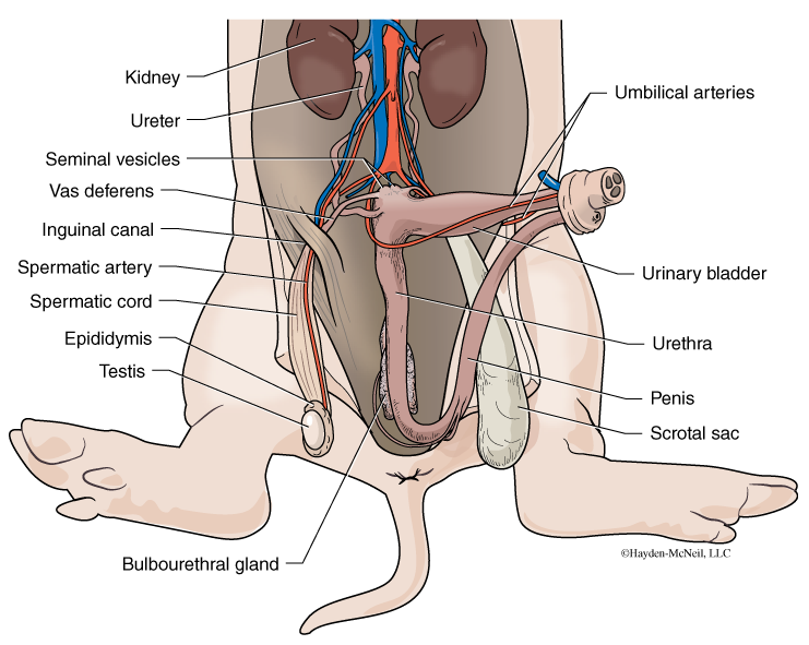
When ejaculation occurs, sperm leave the epididymis and travel through the vas deferens to the urethra. Several additional organs contribute to the ejaculate and are considered accessory reproductive glands. The seminal vesicles, located at the juncture of the vas deferens and urethra, secrete alkaline fluids that neutralize the acidity of the vagina, and they provide fructose that serves as an energy source for the sperm. The bulbourethral gland is caudal to the vas deferens and lies along the side of the urethra. It also produces an alkaline fluid that helps to neutralize the acidity of the vagina. The mixture of sperm and the secretions of the seminal vesicles and bulbourethral glands pass through the urethra that is found within the penis, the structure that deposits the sperm in the female reproductive tract. In the fetal pig, the penis is enclosed by epithelium and is not yet external. These structures are shown in Fig. 5.4.
PROCEDURE
First examine your fetal pig and other pigs in the lab until you are able to consistently identify the sex as well as these external structures: the urogenital opening (males and females), urogenital papilla (female), and the anus (both sexes) and scrotal sac. Look around the room for pigs that may be mature enough for the testes to have descended into the scrotal sac.
If you have a male fetal pig, continue here. If you have a female, skip to the next section. In either case, you must be familiar with both sexes.
Next, use your scissors to extend the incision caudally toward the anus. To see all of the structures, use a scalpel and carefully cut through the pubic symphysis, the bone that is ventral to the anus. It is best to cut away from the midline of the body. Once the region is loosened, use a blunt probe or fine probe to gently loosen the connective tissue or fascia from around the organs you are trying to see. With the male, find the scrotum and with your scissors, open it to see the testis and the epididymis. If the male is not yet mature enough for the testes to have descended, they can be found anterior to a membranous cremasteric pouch. The spermatic cord should be visible on the cranial end of the pouch. The spermatic cord contains the vas deferens, artery and vein, lymphatic vessels and nerves. Carefully slit the cremasteric pouch. Leave the testis and epididymis attached. Using the figure as a guide, follow the spermatic cord cranially and look for where the vas deferens joins the urethra. Find the seminal vesicle and the bulbourethral glands, as well as the urinary bladder and the penis. The urinary bladder lies along the umbilical arteries.
Examine a microscope slide of the cross section of the testis and find these cells: spermatogonium, spermatids, and sperm; find these structures: seminiferous tubules and epididymis.
You should be able to recognize all structures listed here and know their function. Use your text to compare the fetal pig with a human.
IV. FEMALE REPRODUCTIVE SYSTEM AND OOGENESIS
The female reproductive system is equally complex and interesting. The female gonad is the ovary, and it is here that ova (eggs) and several hormones are produced. The ova are produced in a process called oogenesis. There are some very interesting differences between the process of spermatogenesis and oogenesis.
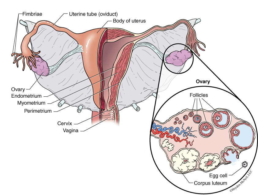
In mammals, oogenesis begins before birth. In a fetus, there are thousands of oogonia present in the fetal ovaries. No new oogonia are produced after birth. Later in the development of the fetus, some of the oogonia may start to grow and become primary oocytes. These primary oocytes will start the process of meiosis but will soon go into a resting state in prophase I. In some cases in humans, this resting state can last for decades. Once puberty starts, several primary oocytes, with the granulosa cells surrounding them, will restart the development and continue through meiosis. The oocyte and granulosa cells together are called a follicle, and it is possible to see follicles at various stages of development in a slide of the cross section of an ovary (see Fig. 5.5).
As the follicle grows, the primary oocyte completes its first meiotic division. However, instead of being a division that produces two identical haploid cells, in oogenesis this first division produces one large cell called the secondary oocyte, and one very small cell called the polar body. Most of the cytoplasm gets concentrated in the secondary oocyte that may eventually become the ovum. The polar body consists mostly of chromosomes. The secondary oocyte starts the second meiotic division, but again gets arrested and will complete that division only if it is fertilized.
In a slide of a cross section of a human ovary, you can see follicles at several different stages of development (Fig. 5.5). As the oocyte develops, it may become separated from the follicle cells by a layer called the zona pellucida, a layer made of glycoproteins. This surface will bind with the sperm if this particular oocyte ever gets fertilized. As maturation continues, the follicle moves closer to the outer edge of the ovary. When ovulation occurs, the follicle ruptures releasing the secondary oocyte. A portion of the follicle remains in the ovary and is called the corpus luteum (literally, “yellow body”), and becomes an endocrine gland that secretes estrogen and progesterone. As long as these hormones are present, the endometrial lining of the uterus is maintained and no additional follicles can develop.
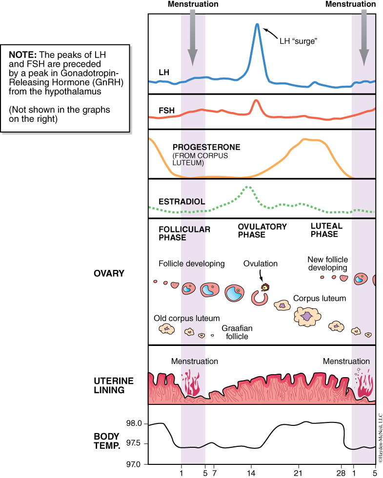
Hormones control the entire process. The maturation of primary oocytes is in response to FHS (follicle stimulating hormone) produced by the anterior pituitary gland in the brain. The follicle cells produce estrogens. Rising levels of estrogen cause an increase in the endometrial lining of the uterus. And the rising estrogen causes the anterior pituitary to release luteinizing hormone (LH) that in turn causes ovulation, or the release of the egg from the follicle in the ovary. You should be familiar with the menstrual cycle in humans (Fig. 5.6) and understand the source for the different hormones.
Once ovulation has occurred, the oocyte is carried into the oviduct by the fimbria (fringe) of the oviduct. The release of hormones at ovulation causes the fimbria to sweep the oocyte into the oviduct. The oviduct is lined with cilia that then moves the oocyte toward the uterus. Fertilization occurs in the oviduct but if it does not occur, the oocyte will degenerate. If fertilization does occur, the blastocyst will implant in the lining of the uterus.
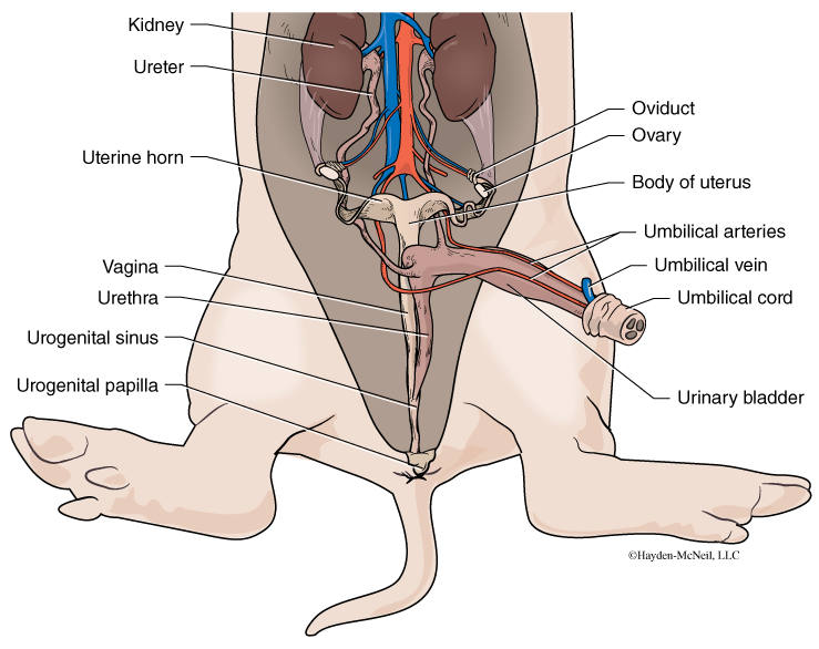
In pigs, the uterus is long and thin and is divided into two sections called uterine horns (see Fig. 5.7). Note that pigs typically have 8–10 piglets in their litter, and current research is finding genetic markers to screen for pigs that could have large litter sizes. The uterine horns join with the vagina at the cervix. The vagina is joined by the urethra and both open to the outside via the urogenital opening. The urogenital papilla is homologous with the penis in the male.
PROCEDURE
As with the male, for the female you will use scissors to extend the incision caudally toward the anus. To see all of the structures, use a scalpel and carefully cut through the pubic symphysis, the bone that is ventral to the anus. It is best to cut away from the midline of the body. Once the region is loosened, use a blunt probe or fine probe to gently loosen the connective tissue or fascia from around the organs you are trying to see. You should first find the ovaries and the oviduct. Follow the oviduct to the uterine horns and uterine body. Next find the vagina and urinary bladder.
Be sure to examine both males and females.
Next, look at a microscope slide of the cross section of an ovary. (A human ovary is about the size of a large almond.) You should be able to identify follicles at various stages of development. You should know all the stages of the human menstrual cycle.
V. EXCRETORY SYSTEM
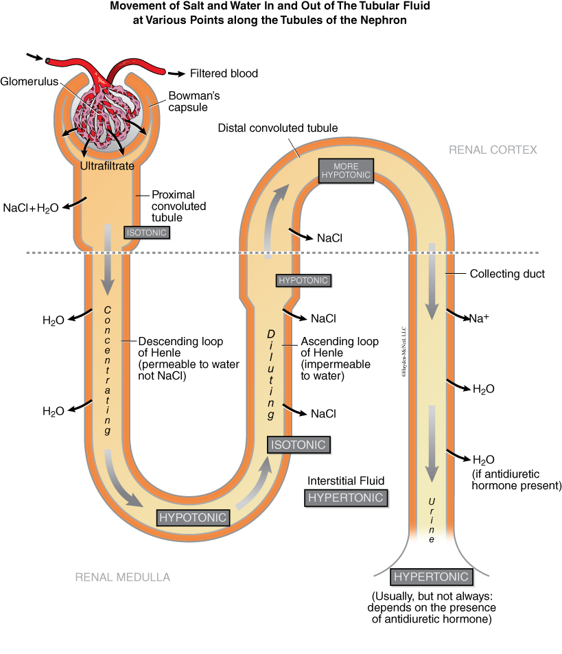
The excretory system is morphologically tightly associated with the reproductive systems, so we will include it here. The excretory system has several functions, including osmoregulation and elimination of nitrogenous wastes. The kidneys filter blood from the circulatory system and have a remarkable ability to concentrate the wastes in the urine. As much as 2000 liters (more than 500 gallons!) of blood are filtered each day. Thankfully, most of the fluid is reabsorbed. In lecture, you will cover the details of urine formation. Chapter 47 in your text, entitled “Osmoregulation and Disposal of Metabolic Wastes,” has a thorough coverage of the topic.
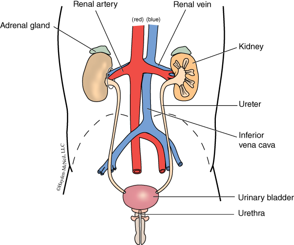
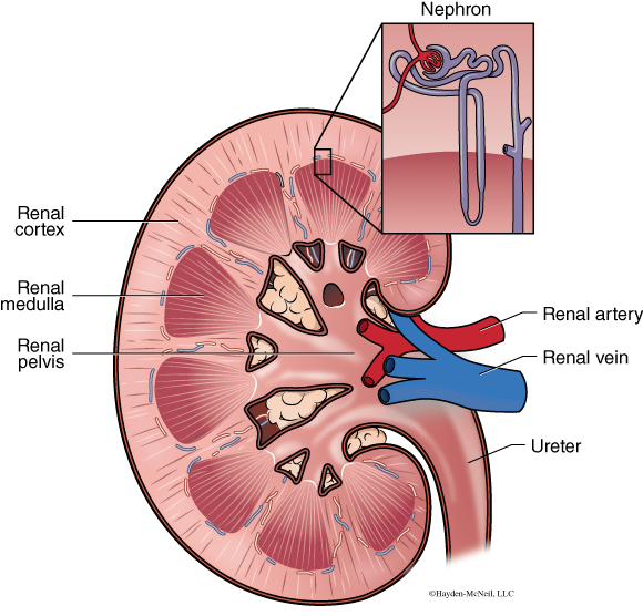
A large artery and vein go to each kidney (Fig. 5.8). The size of the renal artery reflects the importance of blood passing through this organ. The renal artery divides quickly into a capillary bed in the cortex (outer region) of the kidney. The functional unit of the excretory system is the microscopic nephron (Figs. 5.9 and 5.10). Each human kidney has around one million nephrons. One part of the nephron is a knot of capillaries surrounded by a capsule. This knot is called a glomerulus and it is surrounded by Bowman’s capsule. Bowman’s capsule receives fluid (called filtrate) forced out of the glomerulus and carries the filtrate along a proximal convoluted tubule, the loop of Henle and the distal convoluted tubule. The wastes then pass into a collecting duct. Collecting ducts converge to form the renal pelvis. From here the urine passes down the ureter to the urinary bladder, where it is stored before it is eliminated out the urethra.
PROCEDURE
Start by finding one of the kidneys in your fetal pig. Cut the kidney similar to the one in Fig. 5.9 to see the renal pelvis and the connections with the ureter. In addition, you should be able to identify the renal cortex (outermost area) and renal medulla. Find the renal artery and renal vein. Trace the ureter to the urinary bladder and then find the urethra. Be sure to look at both male and female fetal pigs!
Finally, examine the microscope slide of the cross section of the kidney. You should be able to identify the renal cortex and renal medulla on the cross section. In addition, you should be able to identify a glomerulus (capillary bed that forces fluid out of the bloodstream) and Bowman’s capsule (epithelial layer surrounding the glomerulus that receives the filtrate).
VI. NOTES TO INSTRUCTORS AND MATERIALS FOR LAB
Students should be able to recognize male and female fetal pigs both externally and internally. Complete the dissections as indicated. Students should be able to compare and contrast oogenesis and spermatogenesis, as well as recognize cross section of the ovary and testis.
Excretory system: Complete dissection as indicated.