Chapter 9. FUNGI AND ANIMAL DIVERSITY 1
I. OVERVIEW: THE UNIKONTS
What is an animal? What is a fungus? How are these two groups related? Our focus this week is on this important clade, known as the unikonts. The clade is comprised of the amoebozoans (including the Amoeba) and the opisthokonts, which include the animals, the fungi, and the choanoflagellates.
The term unikont refers to the flagellum. In many of these groups, there is a flagellum present at some point in the life cycle of the organism. If present, it is singular. For example, the sperm in animals often have a single flagellum. The term opisthokont refers to a flagellum that is posterior (as in a sperm), as opposed to anterior, as is seen on all other eukaryotes. In addition to the similarity of the flagellum in the opisthokonts, DNA sequences support our understanding of these relationships.
Choanoflagellates are a free-living group that are curiously similar to the cells in sponges.
In this lab we will spend the first half of the class focusing on the fungi, and the second half and next week focusing on animals.
II. FUNGI
Members of the Kingdom Fungi are fascinating and diverse. Fungi may be best known as mushroom or mold spores, but they are actually quite varied and provide very important ecosystem services. In many ways, they are the unsung heroes of the natural world: helping plants to absorb nutrients, breaking down organic material, and making the nutrients available for other organisms. In the Department of Biology at the University of Mississippi, Jason Hoeksema studies the symbiotic association between plant roots and mycorrhizal fungi.
Fungi live by absorptive heterotrophy. They secrete enzymes that break down organic molecules, and they then absorb the nutrients. Many are saprobes, feeding on already dead material; many others are parasites that feed on living material. Still other are mutualists and live symbiotically associated in a way that benefits both partners. Fungi probably arose from a unicellular flagellated protist more than 460 million years ago (the oldest fossils of fungi are from 460 million years ago) and have evolved into over 100,000 species in five clades. The body form varies extensively, from unicellular yeasts to filamentous structures made from branching hyphae. Individuals can range in size from being a single cell, to being a giant underground mycelium that covers miles. (For example, in Oregon, there is a single fungus, living mostly underground, that is found over 2200 acres! This fungus may be over 2500 years old.) Even though it is mostly a terrestrial kingdom and its members grow most easily where conditions are moist, the spores of many fungi can withstand very dry and harsh conditions, making them resilient and among the first to invade an environment.
Fungi are important to people directly in a variety of ways, and are also in important via their effects in the natural world. Fungi are important in decomposition of materials in the natural world, and also in the human structures. They are used extensively in making some foods, for examples, beer and bread, some cheeses, and soy sauce. Your text states that there are 2000 edible species of fungi (with perhaps 5–10 species available locally) and there are around 70 species that are toxic to people, sometimes fatally so. Many plants have symbiotic mycorrhizal fungi associated with their roots and there is evidence that this association dates back 460 million years and reflects when both plants and fungi moved on to dry land. Plants with mycorrhizae grow faster.
In biological research, yeast has been a model eukaryotic cell, and was the first eukaryote to have its genetic code be completely sequenced. Fungi have coexisted with bacteria in many environments for hundreds of millions of years and have probably long competed for some of the same resources. It is no surprise, then, that fungi are an important source of antibiotics (the most famous is penicillin, produced from the fungus genus Penicillium). Penicillin has arguably done more to change the world than any other single compound.
Fungi also cause animal and plant diseases. 70% of all crop diseases, including smuts and rusts, are caused by fungi. The chestnut blight fungus killed most of the native American chestnut trees early in the 20th century, removing a beautiful large tree from the forests of North America. Diseases in humans caused by fungi include ringworm and athlete’s foot, as well as yeast infections that can infect the vagina or the mouth.
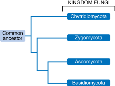
THE CHYTRIDS (CHYTRIDIOMYCOTA)
The chytrids are considered to be the most basal group of fungi. In many ways they are most similar to the protist ancestor, as they still have some flagellated cells. Most other fungi lack flagellated cells, and probably lost the flagellated cells in evolution as they moved on to dry land where flagella could not work. Importantly, some species of chytrids are known to kill large numbers of amphibians and they may be a key piece in the global decline of amphibians.
THE ZYGOMYCETES (ZYGOMYCOTA)
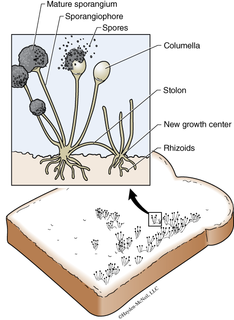
The zygomycetes are best known as bread mold (Fig. 9.2). They show several important characteristics that are generally true of fungi, so we will look at bread mold in some detail. First, they grow by elongating filamentous hyphae, a mass of the hyphae forms a mycelial mat, or a mycelium. The hyphae of zygomycetes are coenocytic; that is, the cells may be multinucleate, without clear divisions or septa between the cells. Spores are microscopic and are produced in the mature sporangia. There can be 50,000 spores in a single sporangium!
Spores are haploid and are a form of asexual reproduction. They are the dispersal phase and move easily through the air. If fact, fungal spores are present just about everywhere. Once a spore lands on a suitable substrate, it will germinate and grow into a new hypha. Hyphae may occur in two different forms or mating types: the “+ form” and the “– form.” Although we cannot see any difference in these two mating types, they can recognize each other chemically, and will grow toward each other. In some cases they will fuse and form two gametangia. These gametangia fuse in a process called plasmogamy, a process that produces one cell with two haploid nuclei. The two nuclei do not fuse at first, but will do so after several divisions. The fusing of the nuclei is called karyogamy, and results in a zygote with a diploid nucleus. The zygote develops into a zygospore. The zygospore may be dormant for a time, and probably undergoes meiosis around the time of germination. The cells that start growing are then haploid and the resulting hypha is haploid as well. Thus the only diploid portion of the life is the zygote and zygospore. What are the advantages of reproducing sexually?
THE ASCOMYCETES (ASCOMYCOTA)
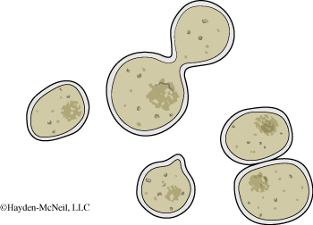
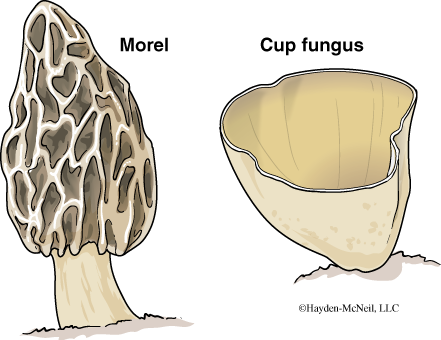
Ascomycetes are a diverse group that appear to be quite different from each other. Sexual spores are called ascospores, which are produced in asci that are found in the ascocarp, or fruiting body. The fruiting body is generally the only part of the life cycle that is visible. An atypical ascomycete are the yeast.
PROCEDURE
Examine the morels and other ascomycetes on the side table. There are also prepared slides for you to look at. How can you recognize an ascomycetes?
THE BASIDIOMCETES (BASIDIOMYCOTA)
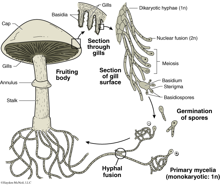
Perhaps the best-known group within the fungi are the Basidiomycota. This group includes some of the mushrooms that we eat as well as shelf fungi and puff balls. Most of the mass of a basidiomycetes is underground. When conditions are right the hyphae will grow into a fruiting body, the mushroom. Here is how it happens: Two hyphae from two mating types will fuse, but the two haploid nuclei from this mating will stay as distinct nuclei in the cell (plasmogamy). The cells will go through mitosis and form a hypha, but the nuclei will remain distinct, creating a dirkaryotic hypha, where each cell has two haploid nuclei. This is denoted as n + n. When conditions are appropriate (typically when there is a lot of moisture), the hypha will grow into a fruiting body that we know as a mushroom (also called a basidiocarp). The stalk and cap of the mushroom are masses of hyphae. The underside of the cap has gills, thin filaments that hold the basidia (or singular basidium), and it is the basidia that produce basidiospores (sexual spores) in the following manner. Karyogamy (or nuclear fusion) on the gills form a diploid zygote, the only portion of the life cycle that is diploid. Meisosis occurs, resulting in four cells, each of which is called a basidiospore.
PROCEDURE
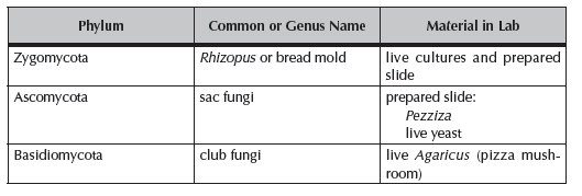
First examine the growth of the Rhizopus in the Petri plate using a dissection microscope. Note the dark sporangia. Can you see individual spores? Note: Keep the lid of the culture closed. Next examine the prepared slides of Rhizopus, and sketch and label hyphae, mycelia, sporangia, and spores.
Examine the morels and other ascomycetes on the side table. There are also prepared slides for you to look at. How can you recognize an ascomycetes?
For the Basidiomycoa, examine a Coprinus, also known as a “pizza mushroom.” Sketch this mushroom. Locate the gills and very gently remove a portion of a gill to examine with a microscope. You should also gently scrape a small part of the stalk or cap to get some tissue to make a wet mount. What are the stalk and cap made from? From the gill, identify the basidiospores.
Critique this statement:
Penicillin has arguably done more to change the world than any other single compound. Why might this be true?
You should know the formal and common names for each of these groups as well as what is in the lab book about each. You should know a representative life cycle of each group.
UNDERSTANDING ANIMAL DIVERSITY
What is an animal? While this question may seem trite if you think of horses and cows and people, it is more interesting when you think about sponges or the jellyfish or even the sea squirts that superficially look like a loosely organized form of jelly but are closely related to the vertebrates.
How do we make sense of the 1.5 million or more species of animals that exist in the world? How do we understand how a starfish might be related to a shark, or how a spider is related to a “daddy longlegs”? How do we understand how the shape of an earthworm helps it to live in the soil or how insects fly? How is it that distantly related groups like dragonflies and bats have each evolved the ability to fly? How is it that the great blue whale, a marine mammal, can be many times bigger than the African elephant, the largest of the land mammals?
There are several tools in the tradition of biology that are helpful in understanding the vast diversity of animal life on earth. In these next two labs, we will explore some of the diversity and some of the means that we have for exploring relationships and adaptations. There are two important principles in diversity. Organisms that are closely related share common derived characteristics. And, organisms that live in the same environment will often share adaptations for that environment, even though they may be only distantly related. Part of what we try to do in understanding diversity is to parse the part that is determined by ancestry and the part that is determined by environment.
In addition, there is a challenge in understanding diversity. You have to know the names and characteristics of some animals to understand how there are similar patterns across taxa and how there are very interesting adaptations seen in just about every species. It requires both some learning material and some integrating what you learn into patterns that are understandable.
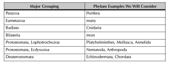
The broadest division is between the Parazoa (literally “alongside animals”) and the Eumetazoa (“true, later animals”). The Parazoa include the sponges, animals that are remarkable different from all of the other animals, all of which are grouped in the Eumetazoa.
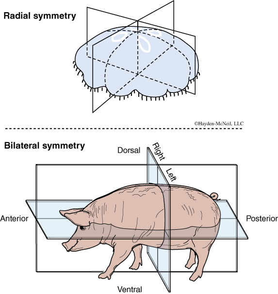
The Eumetazoa are then divided into the Radiata, animals with radial symmetry and the Bilateria, those with bilateral symmetry. The Radiata include the Cnidaria, a phylum we will look at in some detail. In addition to radial symmetry, the Radiata also have only two tissue layers: the endoderm and ectoderm (e.g., they are diploblastic), and if there is a digestive system, it has only one opening.
The Bilateria include most of the rest of the animal kingdom. Members of the Bilateria tend to show cephalization, or the development of a head region, where sensory systems are concentrated and where the nervous system is concentrated. In addition, animals in the Bilateria have three distinct germ layers: ectoderm, endoderm, and mesoderm. They are said to be triploblastic. During development, these germ layers become the different tissues of the body. The digestive system in the Bilateria generally has two openings: the mouth and the anus.
Triploblastic animals are classified on the basis of whether a body cavity is present and on what form the body cavity takes. The word coelom was introduced in the first lab on animal development and it refers to a fluid-filled body cavity. The body cavity is apparently very important in the evolution of larger size in many animals. Animals lacking a body cavity are said to be acoelomate (“no coelom”) and are generally very small. Some animals have a body cavity not lined with mesoderm, called a pseudocoelom. Most animals have a body cavity that is completely lined by mesoderm (mesenteries), and this is called a “true coelom.”
Based on their early development and how the coelom develops, animals can be grouped as either Protostomata or Deuterostomata.
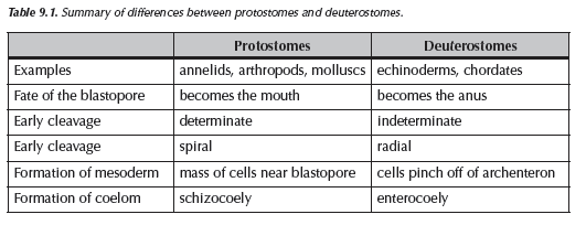
The protostomes are divided into the Lophotrochozoa and the Ecdysozoa. The name Lophotrochozoa is based on the two groups that make up this category, the lophophorates and those with trochozoa larvae. The Ecdysozoa is comprised of animals that go through ecdysis or the molting process. The deuterostomes include the Echinodermata and the Chordata. In lab this week, we will focus on the earliest animals, the protostomes and the Lophotrochozoa. We will emphasize the animals that are less well known.
Be comfortable with these terms and divisions:
Radiata and Bilateria
Acoelomate, Pseudocoelomate, Eucoelomate
Protostomata, Deuterostomata
Lophotrochozoa and Ecdysozoa
III. THE EARLIEST ANIMALS: PHYLUM PORIFERA, THE SPONGES
The Porifera are among the most curious of animals (Fig. 9.6). They have no tissues, no real symmetry, they are not very mobile, and were once considered plants. Although they are mostly marine, there are a few freshwater sponges. For many sponges their shape depends extensively on the environment. If they live in an area without waves, they may grow tall. In areas with waves, they may be much flatter and more encrusting. Their evolutionary history is long. The earliest fossils are from 600 million years ago, and they have been evolving independently of other animals ever since then.
They may be an important source of pharmaceuticals and are being actively investigated by the National Center for Natural Products Research found on the campus of the University of Mississippi. Since sponges are not very mobile, their main defense is by chemicals, and as they have had a long evolutionary history, there has been a lot of time for them to evolve chemicals that would inhibit the growth of competitors.
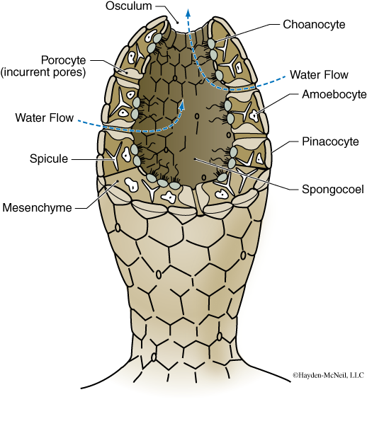
Sponges have specialized cells that help them function. One of these cells is called a collar cell or choanocyte. An ancestor of a sponge may be a Protista similar to the choanoflagellates.
Your TA will review the basic shape of a sponge and how it functions to filter water.
PROCEDURE
Examine the dried sponges available for you on the side table. Note the diversity of shapes and sizes.
Next examine the sponge Grantia on the side table. You will be able to look at a sample with a dissecting microscope. At your own table, examine the prepared slide that is a cross section of Grantia, a small sponge that nicely shows the basic organization of sponges. In your notebook, sketch and label a whole Grantia and the cross section of Grantia. In a short paragraph, describe how water flows through a sponge.
You should understand how a sponge functions and the importance of specialized cells such as the choanocytes.
IV. ANIMALS WITH RADIAL SYMMETRY: THE RADIATA, PHYLUM CNIDARIA
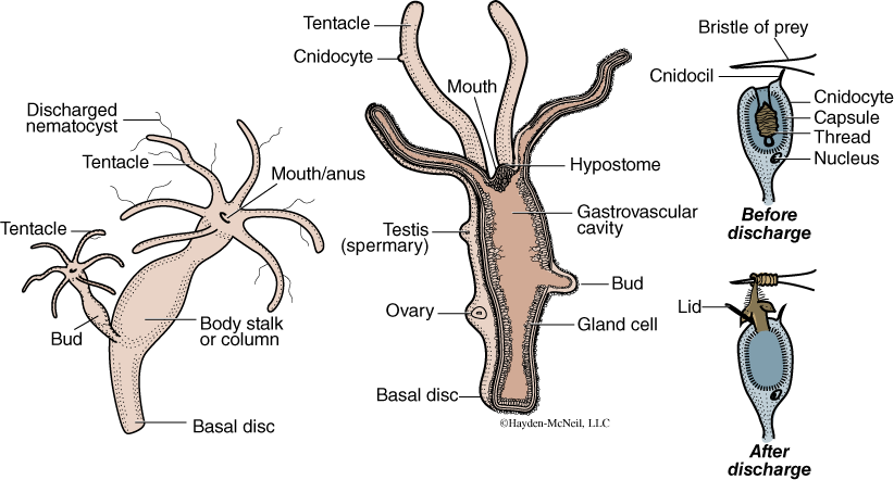
Radial symmetry is one of the forms of symmetry seen in the animal kingdom (Fig. 9.7). One of the interesting and important examples of a group of animals with radial symmetry is the Phylum Cnidaria. This phylum includes many examples of animals that may be familiar to us, including the corals and jellyfish, Hydra, and Portuguese Man ’O War. Radial symmetry means that there are multiple planes of mirror images (Fig. 9.7), compared to the single mirror image seen in bilaterally symmetrical animals.
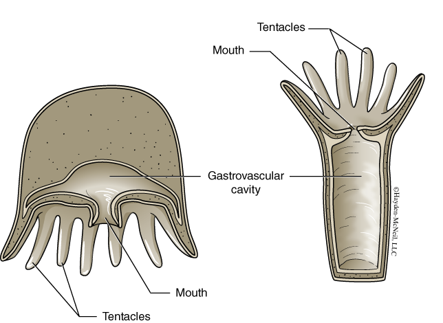
Other important characteristics of this phylum are that individuals have only a single opening to their digestive system (the digestive system is often called a gastrovascular cavity) (Fig. 9.8); their tissues are formed from two germ layers, the endoderm and ectoderm; and they all have cnidocytes (Fig. 9.9), specialized cells for stinging that function in both defense and feeding. Although all Cnidaria have these characteristics in common, they are enormously diverse, with some being very large, others very small, some solitary, some colonial, and some with impressive symbioses.
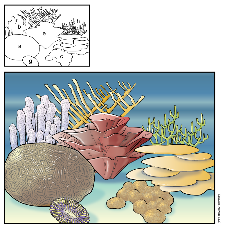
Members of the Cnidaria can occur in two forms: a polyp or medusa. In many cases, both forms are present in the life cycle of a single animal. Coral reefs are made by small coral animals that are polyps. And although the individual animals are very small, over time they can build very large structures (Fig. 9.9).
PROCEDURE
We will start by looking at preserved anemones and jellyfish to understand the difference between the polyp and medusa forms. There will be preserved examples of each on each of the tables in the lab. Make a labeled sketch of each. Label gastrovascular cavity, the endoderm, ectoderm, and tentacles. These are both solitary.
Next we will examine Hydra as important example of a freshwater solitary polyp. Obtain the prepared slide and sketch a Hydra. There will also be live examples for you to see. Hydra can reproduce both sexually and asexually. There will be slides showing both. Note that in Hydra, when a bud forms, it does not stay attached to the parent animal.
We’ll finish by looking at examples of corals.
V. ANIMALS WITH BILATERAL SYMMETRY: THE BILATERIA
The Lophotrochozoa
Phylum Annelida
Phylum Mollusca
THE PLATYHELMINTHES: A BILATERAL, ACOELOMATE GROUP
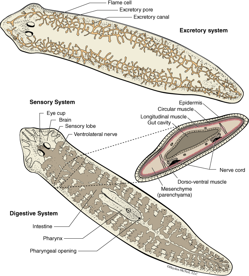
The simplest of the bilateral animals are the flatworms or Platyhelminthes. One well-known example is the free-living planaria, also known as Dugesia (Fig. 9.11). This group lives in freshwater, is small, and has a digestive system with a single opening, known as a pharyngeal opening, that leads to the gastrovascular cavity. Dugesia lacks a body cavity and so, in cross section, the only open space is the gut or the GV cavity.
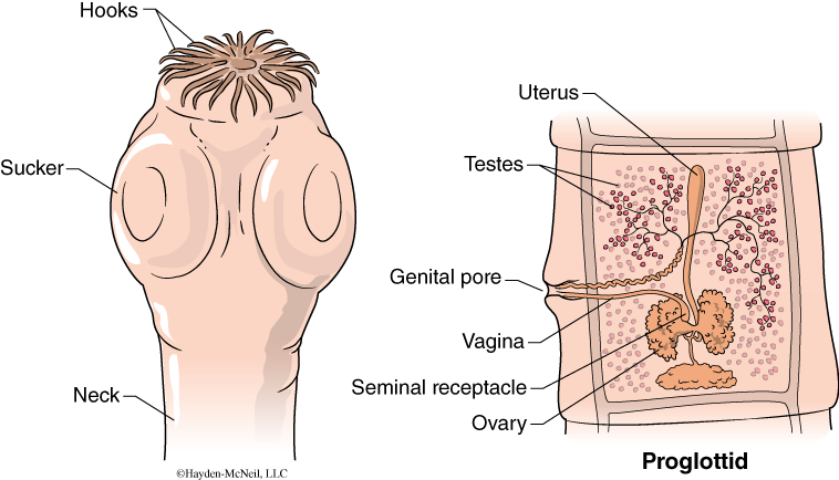
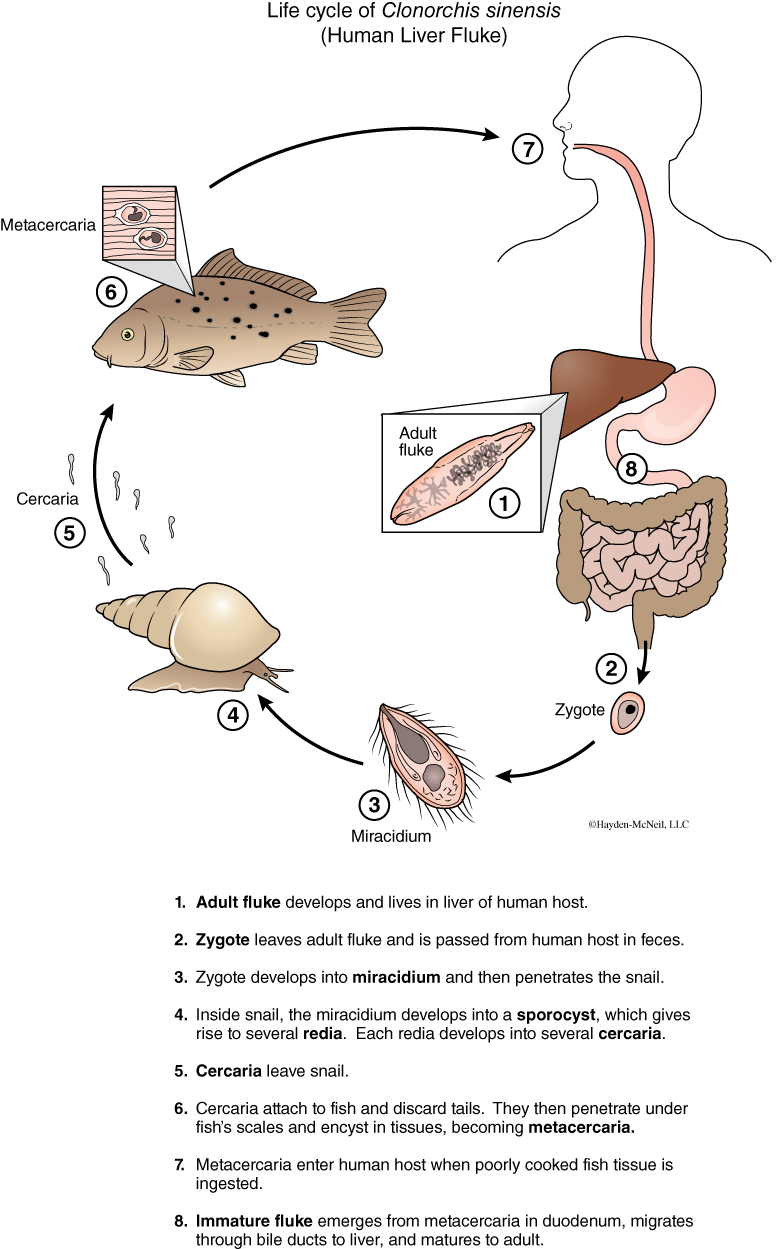
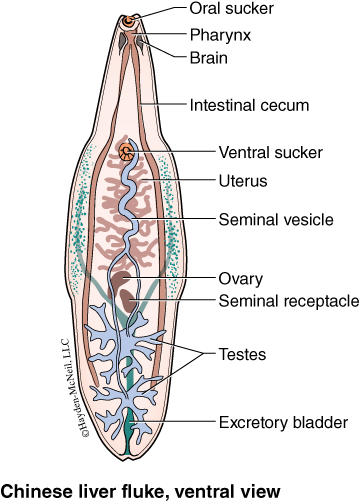
There are also two other groups classified with the Platyhelminthes, but molecular data suggest they may not be closely related to the Planaria. These are both important parasites: the Cestoda or tapeworms (Fig. 9.12) and the Trematoda or flukes (Fig. 9.13 and 9.14). Both of these groups are highly specialized for a parasitic lifestyle. Both lack a coelom and most completely lack a digestive system. They often have an impressively complex life cycle (sometimes with three or four or five different hosts!). And, as is seen in most parasites, much of their body is devoted to reproduction. For example, in the tapeworms, there is no digestive system at all, and most of the internal organs are ovaries.
PROCEDURE
Examine the live Dugesia on the side table.
Next, examine and sketch the whole mount and cross section of the Dugesia. For each cross section, show where on your whole mount that section is taken from.
Get a prepared slide of liver fluke and tapeworm. Make a sketch of each. And be able to recognize each.
PROTOSTOMES, LOPHOTROCHOZOANS; THE ANNELIDA AND THE MOLLUSCA
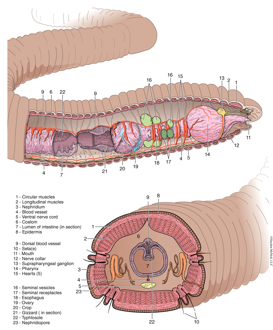
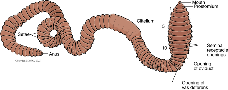
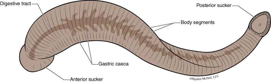
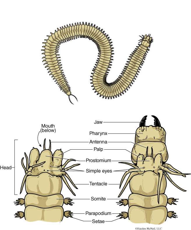
The Phylum Annelida includes the well-known earthworms (Fig. 9.15) and leeches (Fig. 9.16) and a wide variety of lesser-known marine such as the Nereis (Fig. 9.17). Annelids have bilateral symmetry, a true coelom, a tube-within-a-tube digestive system. They also have a closed circulatory system. In addition to the above, they are characterized by being segmented, a characteristic that is visible both externally and internally. This phylum is ecologically important. We will examine examples from the three major groups within the Annelida.
PROCEDURE
We will start with live earthworms and examine how they are able to move. With the live animal in hand, you should look for the external characteristics shown in the figures. Which end is anterior? Which side is dorsal?
Next, look at the cross section microscope slide to get familiar with some of the internal organs. A demonstration dissection will also be available for you to examine.
Compare the earthworm with other members in the phylum: the leech and the marine worm in the genus Nereis. How are the similar?
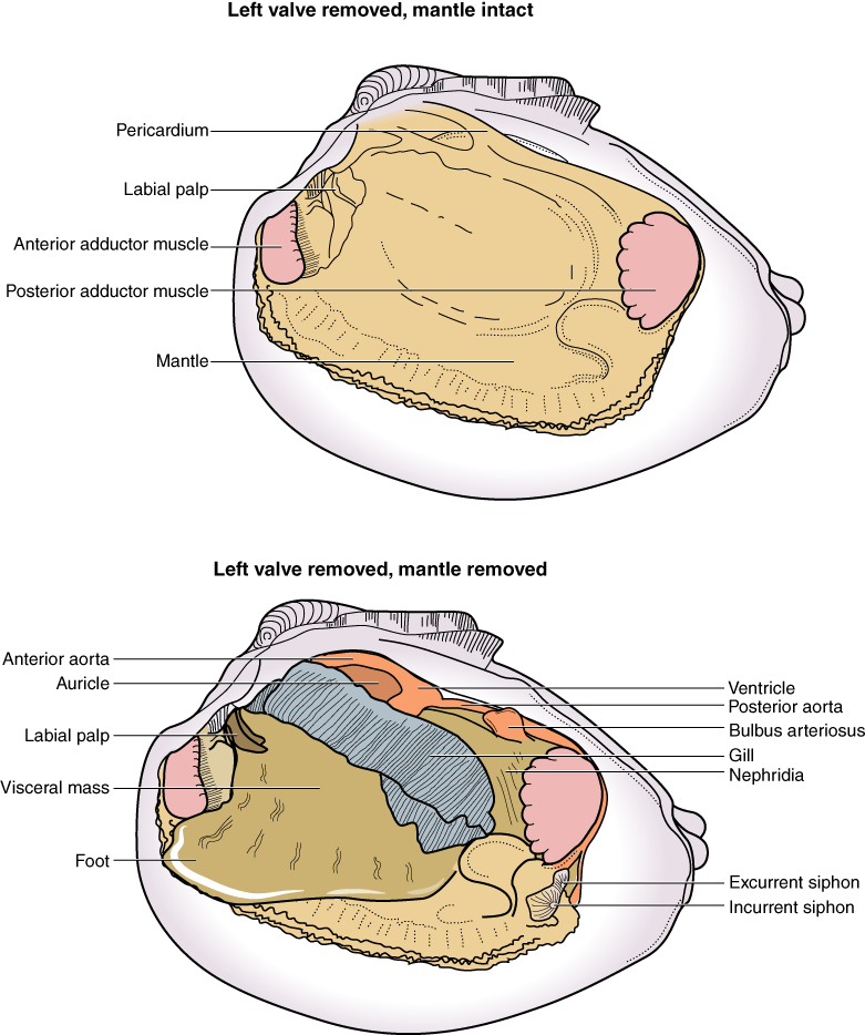
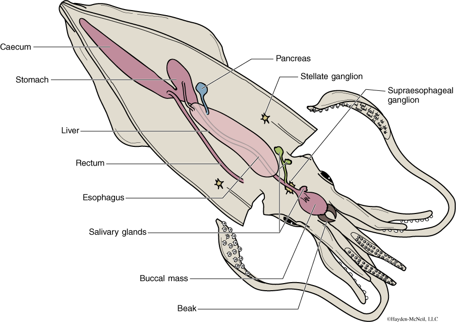
THE MOLLUSCA
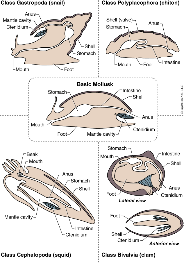
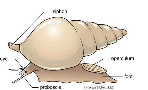
The Mollusca is large group that includes both large and small animals. It includes the giant squid as well as clams and snails. These animals are bilateral protostomes, with a tube-within-a-tube digestive system. While superficially these animals may look very different, there is a variety of traits that they share (Fig. 9.18). They all have a soft body, often covered by a shell. There is a broad muscular foot used for locomotion. This foot takes a variety of forms. There is a visceral mass, located above the foot. And there is a mantle, a layer of cells that secrete the shell. We will look at three examples of Mollusca: the gastropods (snails), the bivalves (clams), and cephalopods (squids).
PROCEDURE
We will have representative examples of three classes in the Mollusca. Sketch an example of each group. For each of the groups, be able to find the foot, the shell (if present), and the visceral mass. Be able to explain the basis for grouping these three classes together.
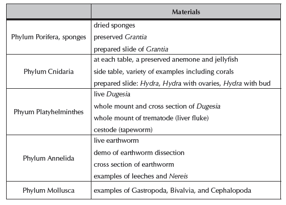
VI. NOTES TO INSTRUCTORS AND MATERIALS FOR LAB