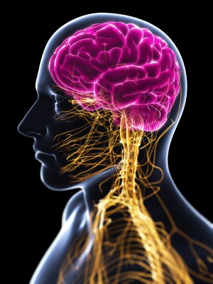

The Central Nervous System: Spotlight on the Brain
The nervous system does its work in all parts of the body. The peripheral nervous system sends signals from the brain to your extremities. It regulates digestion, breathing, and sexual arousal. The peripheral nervous system can be divided into two main categories of function.
There is the somatic system, which we have control over. This includes voluntary movement of muscles. Then there is the autonomic system. The "auto" refers to the fact that this system is self regulated is largely out of reach of conscious control.
Autonomic functions are broken down into sympathetic responses, which are arousing, and parasympathetic responses, which are calming . If one is frightened, a series of sympathetic fight or flight responses happen automatically. Your heart beats faster. Muscles clench. This is the unconscious peripheral nervous system at work.
All that information from the periphery, and from your organs, as well—information about sensation, is your stomach full or not—all that goes to the spinal cord, which is the beginning of the central nervous system.
The spinal cord often orchestrates motor responses without any input from the brain at all. The spinal cord governs our reflexes, which are our automatic responses to stimuli. The pain withdrawal reflex is the result of sensory neurons in the finger sending signals to the spinal cord, where motor neurons signal the muscles to react, withdrawing from the painful stimulus.
As we travel up the spinal cord to the brain, we are confronted with a wonderfully complex structure—the human brain. And when we look inside, the structures we see and the elegant functions they perform, shaped by millions of years of evolution, display the staggering complexity that makes us who we are.
If you follow the spinal cord up into the brain, you will reach the hind brain, the boiler room of the central nervous system. It controls the most basic functions of the body—respiration, alertness, and motor skills. One of the most prominent structures of the hind brain is the cerebellum, which is Latin for "little brain"—rightly so, as this structure, with its characteristic folds, sits tucked beneath the cortex it resembles above and the brain stem to one side.
The cerebellum is a spongy-like structure in the back of the head. And it plays a very important role in governing the fine motor skills that everyone possesses. And in particular, that artists possess when musicians play the piano or an ice figure skater skates his or her routine on the ice—a lot of the delicate fine motor movements that you need to make are controlled by the cerebellum.
Above the cerebellum, the forebrain is divided into two main regions—the cerebral cortex and the subcortical structures, which themselves are divided down the middle into two hemispheres. The subcortical structures are closest to the mid-brain structures atop the brain stem, and they are evolutionarily older than the cerebral cortex that wraps around them.
One of the most prominent subcortical structures is the thalamus. Like a massive switchboard, the thalamus sits near the center of the brain, receiving input from all senses, except for smell, and routing them to various parts of the cortex.
And hence, it's in a great position to modulate information and to gate information when needed.
The other subcortical structures—the hypothalamus, the pituitary gland, amygdala, and hippocampus—make up the limbic system. The limbic system is involved in motivation, emotion, learning, and memory. The hypothalamus regulates our most vital internal systems, including hunger and sexual behavior.
It sends signals to the pituitary gland, the conductor of a body's hormone producing system, to release key signals that play a role in stress, hunger, and some of the major physiological transformations of the body, such as breastfeeding.
Well, the pituitary hypothalamic system is one way that your brain monitors what's going on your body and communicates with it—a chemical registration of what's going on with your hormones.
The hippocampus is the seat of new memory formation in the brain.
So the hippocampus is actually one of my favorite areas of the brain. But its functions are fairly well understood. Number one, it's very important for memory formation.
Raw sensory data is held transiently in the brain. Without the hippocampus, the assembly line of memory, we could never convert this raw data into lasting memories.
The other, second function that the hippocampus plays—and this is more well understood in animal research—is that it acts like a GPS. It contains a map of the environment. The activity in the hippocampus will track your position in large scale space.
Information is processed along the assembly line of the hippocampus a certain features are emphasized and others discarded, then stored indefinitely in parts of the cortex for retrieval, sometimes throughout an entire lifetime.
But how do we attach emotional significance to these memories? The answer seems to lie in an almond-sized region called the amygdala. The amygdala is involved in rage, aggression, and fear. The amygdala works to link these experiences and emotions to the processing of emotional memories and to the autonomic bodily reactions that prepares the fight or flight response.
Here, incoming sensory stimuli are stamped with the emotional responses of the body, and lasting memories are formed.
So the amygdala are two almond-shaped structures, again in the middle of the brain. They are well defined, and they are very well understood, thanks to some beautiful work by Joe LeDoux and many other people.
The amygdala is very important, because of its connectivity. It receives information from all of the sensory structures, all of the sensory pathways—vision, auditions, sights, sounds, smells, tastes. And it's able to integrate all those.
Your amygdala might say to you, hey, this is dangerous. You better remember it. The other stuff isn't. You can forget about it, because you don't want to hang onto everything. So it'll overvalue it and tell you to remember.
Surrounding the subcortical structures is the cerebral cortex. This is the wrinkled surface one sees when looking at the outside of the brain with the naked eye. It is the highest level of processing in the brain, and is responsible for the most complex aspects of perception, emotion, movement, and thought.
The folds of the cortex are a feat of evolution. Stretched out, the surface of the cortex is about the size of a newspaper page. But to fit that amount of tissue inside the human skull, the cortex is steadily folded onto itself to increase surface area over evolutionary history within a confined space.
The cerebral cortex is divided into two hemispheres, with each hemisphere more or less symmetrical in their appearance and, to an extent, their functions. The hemispheres are linked by a dense highway of axons in the center of the brain called the corpus callosum. Each hemisphere controls movement in the opposite side of your body, meaning that the left hemisphere controls movement in your right hand. This is called contralateral processing.
So the cerebral cortex has these four lobes. The one all the way in the back is the occipital lobe. And then just in front of it, behind your ear, is the temporal lobe. Above that is the parietal lobe. And then all the way in the front is the frontal lobe.
The occipital lobe, on the back of the head, is one of the more well understood brain areas, because its functions are somewhat more straightforward than other parts of the brain. So the occipital lobe contains visual cortex, where it basically possesses a map of the visual world as it comes through the eyes, gets processed and mapped onto the back of your head, and elementary features of the visual world become analyzed, such that you can start to recognize objects based on that information.
Moving on to the temporal lobe, the visual functions actually continue from the occipital into the bottom parts of the temporal lobe. In addition, in the temporal lobe are areas involved in auditory processing—the sounds that we hear in our daily life, but also the speech sounds that we receive and process and are able to understand.
And the top part of the temporal lobe is the auditory cortex. And the bottom part is the continuation of the visual cortex.
The parietal lobe processes information about touch in an area called the somatosensory cortex, where each area of the body is represented.
It doesn't represent that in a random way, but actually contains a map. It's systematic, such that two adjacent areas in somatosensory cortex will map onto two adjacent areas on your body.
Lastly, the frontal lobe, which sits just behind the forehead, is involved with speech and movement. It contains the motor cortex. The motor cortex send signals out to the body. The motor cortex sense signals so a subcortical structure called the basal ganglia and the cerebellum, then onward to the spinal cord to initiate movements.
The frontal lobe also deals with many of the qualities that make us most human—abstract thinking, planning, executive control, judgment. Throughout all brain structures, association areas are what makes the brain so powerful—the ability to link concepts together to form new ideas and to represent the outside world with such richness on the inside.
This is accomplished by neurons speaking to one another throughout the lobes, across the hemispheres, and across the subcortical and hind brain structures.
These structures are intertwined with one another in processing information. And we shouldn't think of a thought as being located here and an emotion here. These things are circling around and feeding on one another. And they're broad networks. And there's activity throughout this network.
One thought to be a fixed network with a permanent cell population, it turns out that the brain is remarkably plastic, with new neurons being formed throughout a lifetime and old pathways subject to rewiring. The brain is the crown each of us wears, one formed by millions of years of evolution and billions of neurons playing their parts.
So one way to maybe approach this very, very complicated problem is to begin to think more about how systems interact, because we aren't our thoughts, our memories, our, emotions, our motivations. We're all of those things together, and too much research is focused on specific systems and not enough on how the systems really come together.
We need a theory of the brain, and to do that, we have to begin to work on more about how individual systems interact. And I think out of that will come a theory of the brain once we've advanced far enough.