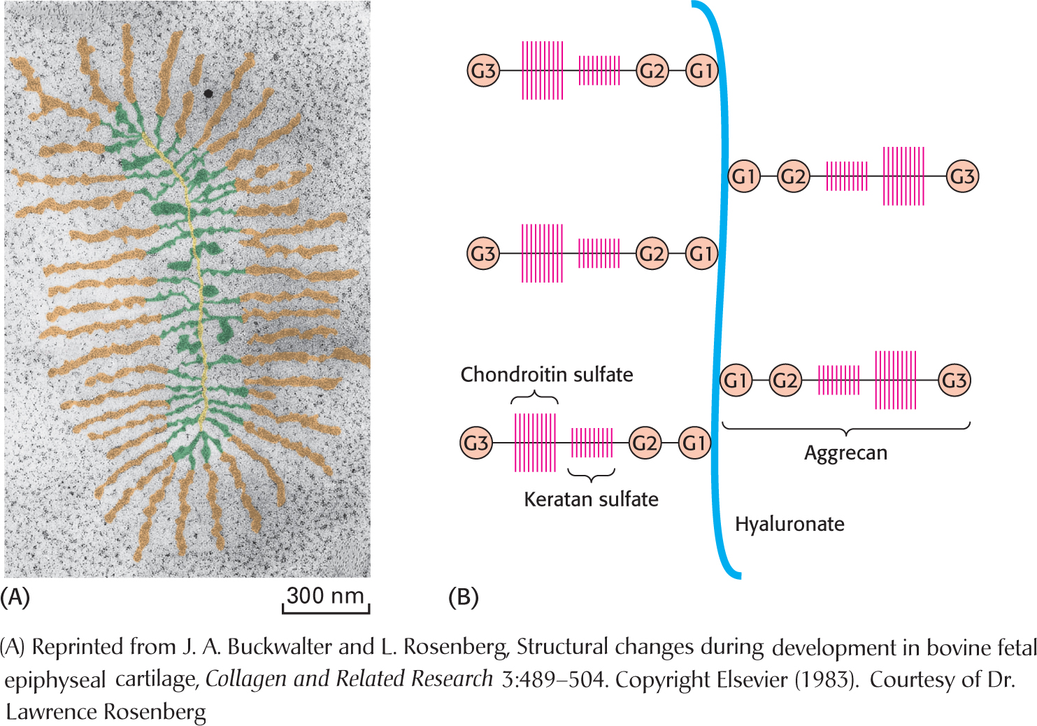
Figure 10.21 The structure of proteoglycan from cartilage. (A) Electron micrograph of a proteoglycan from cartilage (with color added). Proteoglycan monomers emerge laterally at regular intervals from opposite sides of a central filament of hyaluronan. (B) Schematic representation in which G stands for globular domain.