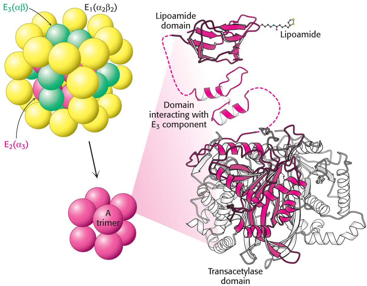
Figure 18.6 A schematic representation of the pyruvate dehydrogenase complex. The transacetylase core (E2) is shown in red, the pyruvate dehydrogenase component (E1) in yellow, and the dihydrolipoyl dehydrogenase (E3) in green. The number and type of subunits of each enzyme are given parenthetically. Each red ball represents a trimer of three E2 subunits. Notice that each subunit consists of three domains: a lipoamide-binding domain, a small domain for interaction with E3, and a large transacetylase catalytic domain. The transacetylase domain has three subunits, with one subunit depicted in red and the other two in white in the ribbon representation.
[Leave] [Close]