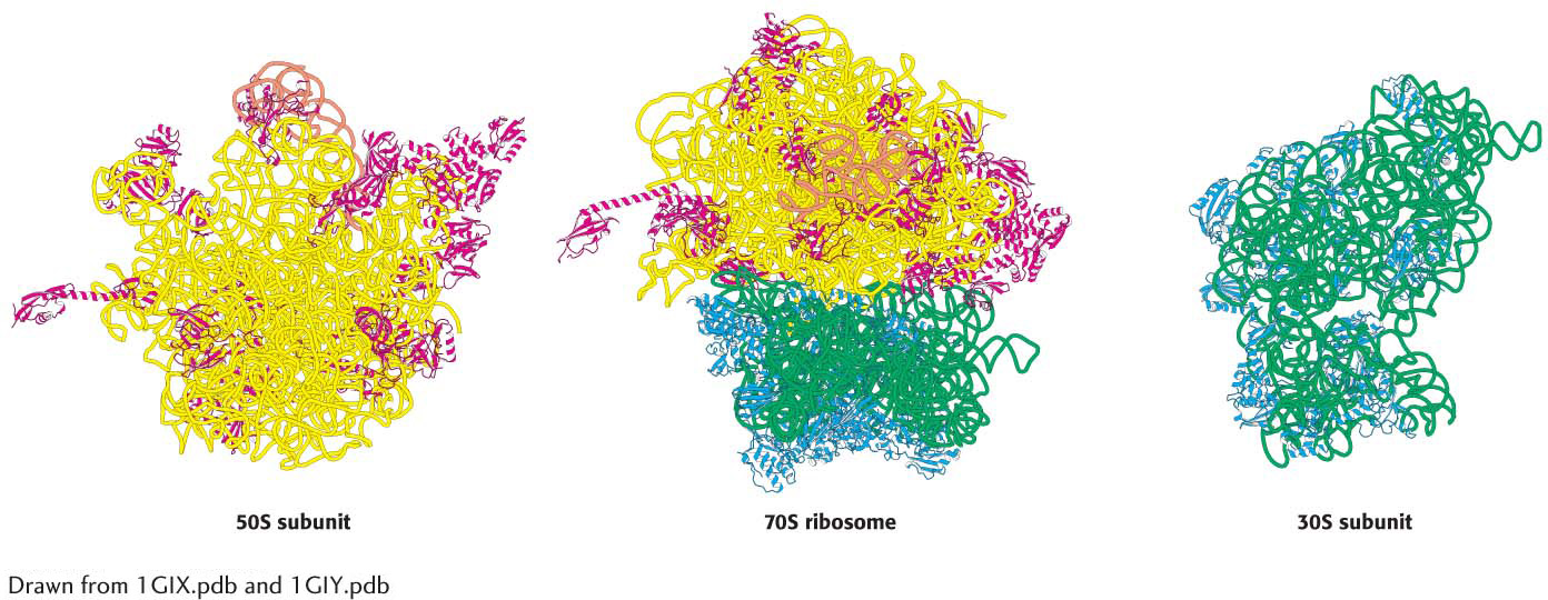
Figure 39.7  Figure 39.7 The ribosome at high resolution. Detailed models of the ribosome based on the results of x-
 Figure 39.7 The ribosome at high resolution. Detailed models of the ribosome based on the results of x-