Figure 4.30 
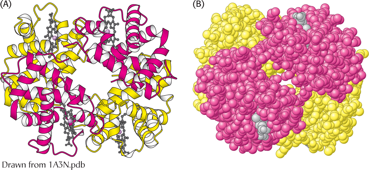
 The α2β2 tetramer of human hemoglobin. The structure of the two identical α subunits (red) and the two identical β subunits (yellow). (A) The ribbon diagram shows that they are composed mainly of α helices. (B) the space-
filling model illustrates the close packing of the atoms and shows that the heme groups (gray) occupy crevices in the protein.

 The α2β2 tetramer of human hemoglobin. The structure of the two identical α subunits (red) and the two identical β subunits (yellow). (A) The ribbon diagram shows that they are composed mainly of α helices. (B) the space-
[Leave] [Close]