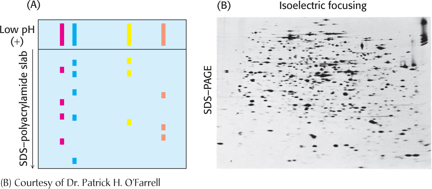
Figure 5.11 Two-dimensional gel electrophoresis. (A) A protein sample is initially fractionated in one direction by isoelectric focusing as described in Figure 5.10. The isoelectric-focusing gel is then attached to an SDS–polyacrylamide gel, and electrophoresis is performed in the second direction, perpendicular to the original separation. Proteins with the same pI value are now separated on the basis of mass. (B) Proteins from E. coli were separated by two-dimensional gel electrophoresis, resolving more than a thousand different proteins. The proteins were first separated according to their isoelectric ph in the horizontal direction and then by their apparent mass in the vertical direction.