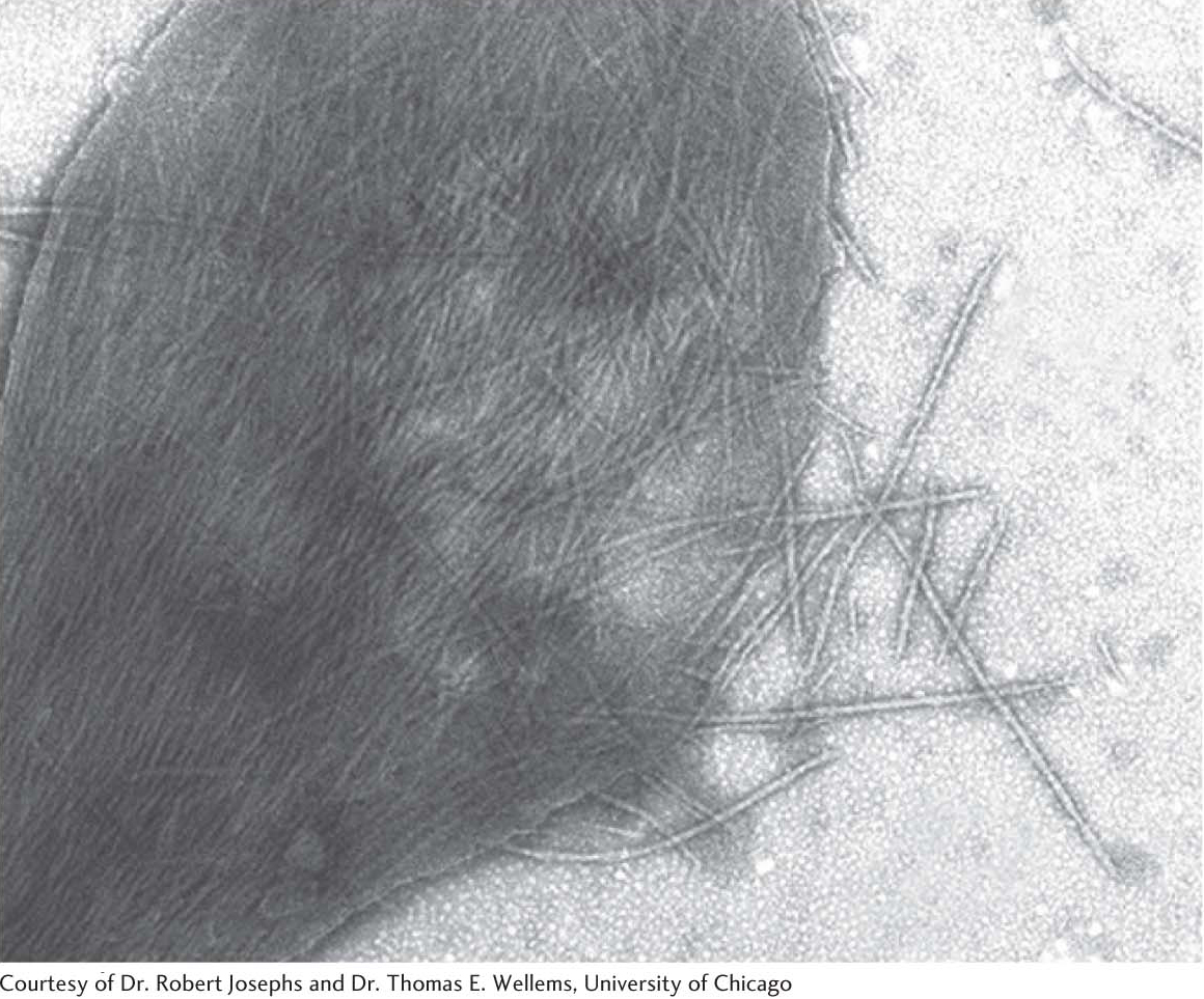
Figure 9.19
Sickl
e-
cell hemoglobin fibers
. An electron micrograph depicting a ruptured sickled red blood cell with fibers of sickl
e-
cell hemoglobin emerging.
[
Leave
] [
Close
]
Next