10.4 Lectins Are Specific Carbohydrate-Binding Proteins

The diversity and complexity of the carbohydrate units and the variety of ways in which they can be joined in oligosaccharides and polysaccharides suggest that they are functionally important. Nature does not construct complex patterns when simple ones suffice. So why all this intricacy and diversity? It is now clear that these carbohydrate structures are the recognition sites for a special class of proteins. Such proteins, termed glycan-binding proteins, bind specific carbohydrate structures on neighboring cell surfaces. Originally discovered in plants, glycan-binding proteins are ubiquitous, and no living organisms have been found that lack these key proteins. We will focus on a particular class of glycan-binding proteins termed lectins (from Latin legere, “to select”). The interaction of lectins with their carbohydrate partners is another example of carbohydrates being information-rich molecules that guide many biological processes. The diverse carbohydrate structures displayed on cell surfaces are well suited to serving as sites of interaction between cells and their environments. Interestingly, the partners for lectin binding are often the carbohydrate moiety of glycoproteins.

183

Lectins Promote Interactions Between Cells

Cell–cell contact is a vital interaction in a host of biochemical functions, ranging from building a tissue from isolated cells to facilitating the transmission of information. The chief function of lectins is to facilitate cell–cell contact. A lectin usually contains two or more carbohydrate-binding sites. The lectins on the surface of one cell interact with arrays of carbohydrates displayed on the surface of another cell. Lectins and carbohydrates are linked by a number of weak noncovalent interactions that ensure specificity yet permit unlinking as needed.

We have already been introduced to a lectin obliquely. Recall that, in I-cell disease, lysosomal enzymes lack the appropriate mannose 6-phosphate, a molecule that directs the enzymes to the lysosome. Under normal circumstance, the mannose 6-phosphate receptor, a lectin, binds the enzymes in the Golgi apparatus and directs them to the lysosome.

Figure 10.26: Selectins mediate cell–cell interactions. The scanning electron micrograph shows lymphocytes adhering to the endothelial lining of a lymph node. The L selectins on the lymphocyte surface bind specifically to carbohydrates on the lining of the lymph-node vessels.

!clinic! CLINICAL INSIGHT: Lectins Facilitate Embryonic Development

One class of lectins consists of proteins termed selectins, which bind immune
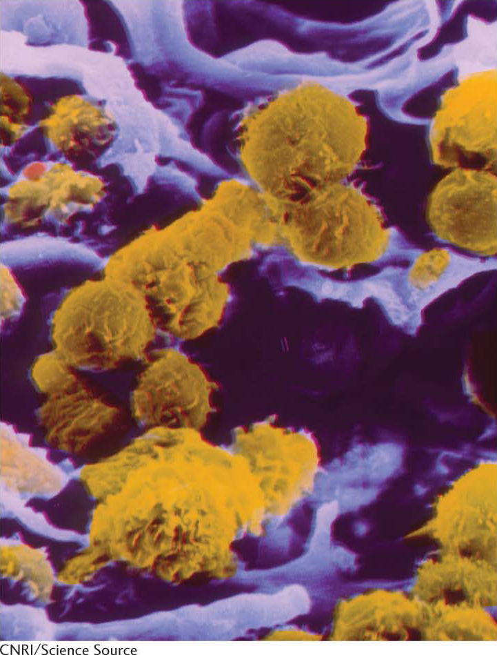
-system cells to sites of injury in the inflammatory response (Figure 10.26). The L, E, and P forms of selectins bind specifically to carbohydrates on lymph-node vessels, endothelium, or activated blood platelets, respectively. L-Selectin, originally thought to participate only in the immune response, is produced by an embryo when it is ready to attach to the endometrium of the mother’s uterus. For a short length of time, the endometrial cells present an oligosaccharide on the cell surface. When the embryo attaches through lectins, the attachment activates signal pathways in the endometrium to make implantation of the embryo possible.

!clinic! CLINICAL INSIGHT: Influenza Virus Binds to Sialic Acid Residues

Many pathogens gain entry into specific host cells by adhering to cell-surface carbohydrates. For example, influenza virus recognizes sialic acid residues linked to galactose residues that are present on cell-surface glycoproteins. The viral protein that binds to these sugars is called hemagglutinin (Figure 10.27A).

Figure 10.27: Viral receptors. (A) Influenza virus targets cells by binding to sialic acid residues located at the termini of oligosaccharides present on cell-surface glycoproteins and glycolipids. These carbohydrates are bound by hemagglutinin, one of the major proteins expressed on the surface of the virus. (B) When viral replication is complete and the viral particle buds from the cell, the other major viral-surface protein, neuraminidase cleaves oligosaccharide chains to release the viral particle.

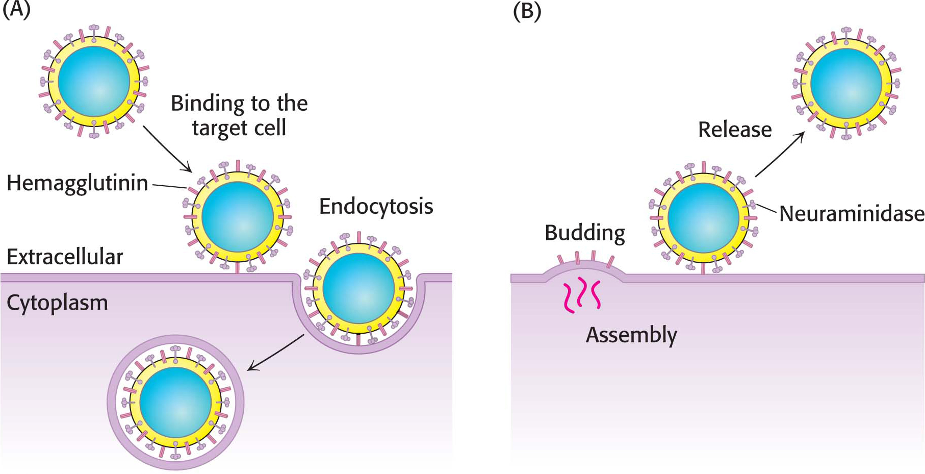
After binding to hemagglutinin, the virus is engulfed by the cell and begins to replicate. To exit the cell, a process essentially the reverse of viral entry occurs (Figure 10.27B). Viral assembly results in the budding of the viral particle from the cell. Upon complete assembly, the viral particle is still attached to sialic acid residues of the cell membrane by hemagglutinin on the surface of the new virions. Another viral protein, neuraminidase (sialidase), cleaves the glycosidic bonds between the sialic acid residues and the rest of the cellular glycoprotein, freeing the virus to infect new cells, spreading the infection throughout the respiratory tract. Inhibitors of this enzyme such as oseltamivir (Tamiflu) and zanamivir (Relenza) are important anti-influenza agents.

184

Viral hemagglutinin’s carbohydrate-binding specificity may play an important role in species specificity of infection and ease of transmission. For instance, avian influenza H5N1 (bird flu) is especially lethal and is readily spread from bird to bird. Although human beings can be infected by this virus, infection is rare and human-to-human transmission is rarer still. The biochemical basis of these characteristics is that the avian-virus hemagglutinin recognizes a different carbohydrate sequence from that recognized in human influenza. Although human beings have this sequence, it is located deep in the lungs. Infection is thus difficult, and, when it does occur, the avian virus is not readily transmitted by sneezing or coughing.