13.2 Receptor Proteins Transmit Information into the Cell

✓ 5 Differentiate the various types of membrane receptors.

Most receptor proteins that convey environmental information into the cell interior fall into three classes: seven-transmembrane-helix receptors, dimeric receptors that recruit protein kinases, and dimeric receptors that are protein kinases. We begin by considering the largest class of receptors, the seven-transmembrane-helix receptors.

Seven-Transmembrane-Helix Receptors Change Conformation in Response to Ligand Binding and Activate G Proteins

Table 13.1 Biological functions mediated by 7TM receptors
Source: Data from J. S. Gutkind, J. Biol. Chem. 273:1839–1842, 1998.

The seven-transmembrane-helix (7TM) receptors transmit information initiated by signals as diverse as photons, odorants, tastants, hormones, and neurotransmitters (Table 13.1). Several thousand such receptors are known, and the list continues to grow. Indeed, approximately 50% of the drugs that we use alter receptors of this class. Mutations in these receptors and their associated components cause a host of diseases, some of which are listed in Table 13.2. As the name indicates, these receptors contain seven helices that span the membrane bilayer (Figure 13.3). An example of a 7TM receptor that responds to chemical signals is the β-adrenergic receptor. This protein binds epinephrine (also called adrenaline), a hormone responsible for the “fight or flight” response. We will address the biochemical roles of this hormone in more detail later (Chapters 16 and 24).

Table 13.2 Diseases resulting from defects in 7TM receptors
Figure 13.3: Figure 13.3 The structure of 7TM receptors. (A) Schematic representation of a 7TM receptor showing how it passes through the membrane seven times. (B) Three-dimensional structure of a subtype (β2) of the β-adrenergic receptor, a 7TM receptor taking part in fuel mobilization. Notice the ligand-binding site near the extracellular surface.

228

The 7TM receptors undergo a change in conformation in response to ligand binding. The binding of a ligand on the outside the cell induces a conformational change in the 7TM receptor that can be detected inside the cell. As we shall see, 7TM receptors also have in common the next step in their signal-transduction cascades.

Ligand Binding to 7TM Receptors Leads to the Activation of G Proteins

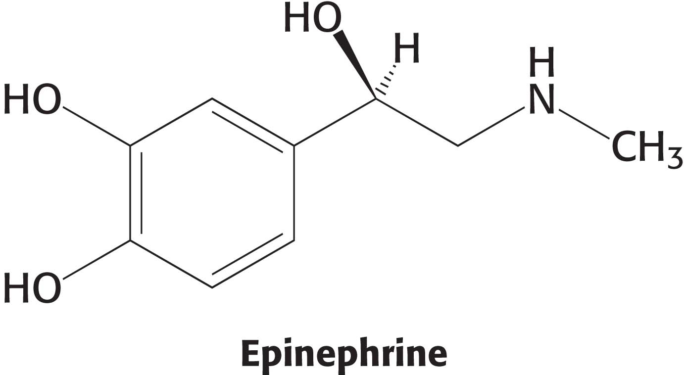
Let us focus on the β-adrenergic receptor as a model of the 7TM receptor class. What is the next step in the pathway after the binding of epinephrine by the β-adrenergic receptor? The conformational change in the cytoplasmic domain of the receptor activates a GTP-binding protein. This signal-coupling protein is termed a G protein (G for guanyl nucleotide). The activated G protein stimulates the activity of adenylate cyclase, an enzyme that increases the concentration of the second messenger cAMP by forming it from ATP (Figure 13.4).

Figure 13.4: The activation of protein kinase A by a G-protein pathway. Hormone binding to a 7TM receptor initiates a signal-transduction pathway that acts through a G protein and cAMP to activate protein kinase A.

How do these G proteins operate? In the unactivated state, the guanyl nucleotide bound to the G protein is GDP. In this form, the G protein exists as a heterotrimer consisting of α, β, and γ subunits; the a subunit (referred to as Gα) binds the nucleotide (Figure 13.5). The α and γ subunits are usually anchored to the membrane by covalently attached fatty acids. The exchange of the bound GDP for GTP is catalyzed by the ligand-bound receptor. The ligand–receptor complex interacts with the heterotrimeric G protein and opens the nucleotide-binding site so that GDP (guanosine diphosphate) can depart and GTP (guanosine triphosphate) can bind. The α subunit simultaneously dissociates from the βγ dimer (Gβγ; Figure 13.4). The dissociation of the G-protein heterotrimer into Gα and Gβγ units transmits the signal that the receptor has bound its ligand. The human genome encodes 15 genes for Gα, one of which (Gαs) is expressed ubiquitously while the others are expressed in specific cell-types.

Figure 13.5: A heterotrimeric G protein. (A) A ribbon diagram shows the relation between the three subunits. In this complex, the α subunit (gray and purple) is bound to GDP. Notice that GDP is bound in a pocket close to the surface at which the α subunit interacts with the βγ dimer (the β subunit is shown in blue and γ in yellow). (B) A schematic representation of the heterotrimeric G protein.

A single ligand–receptor complex can stimulate nucleotide exchange in many G-protein heterotrimers. Thus, hundreds of Gα molecules are converted to their GTP-bound forms from their GDP-bound forms for each bound molecule of hormone, giving an amplified response. All 7TM receptors appear to be coupled to G proteins, and so the 7TM receptors are sometimes referred to as G-protein-coupled receptors or GPCRs.

229

Activated G Proteins Transmit Signals by Binding to Other Proteins

As just described, the formation of the ligand–receptor complex activates a G protein. How does the G protein propagate the message that the ligand is present? It does so by a variety of means, depending on the specific type of G protein. We will begin by examining one target of a G protein, the enzyme adenylate cyclase (Figure 13.6). The adenylate cyclase enzyme that is activated by the β-adrenergic signaling pathway is a membrane protein that contains 12 presumed membrane-spanning helices. The Gα protein binds to adenylate cyclase on the Gα surface that had bound the βγ dimer when the Gα protein was in its GDP form. Gαs (“s” stands for “stimulatory”) stimulates adenylate cyclase activity, thus increasing cAMP production. The net result is that the binding of epinephrine to the receptor on the cell surface increases the rate of cAMP production inside the cell.

Figure 13.6: Adenylate cyclase activation. (A) Adenylate cyclase is a membrane protein with two large intracellular domains (red and orange) that contain the catalytic apparatus. (B) The structure of the complex between Gα in its GTP form bound to a catalytic fragment of adenylate cyclase. Notice that the surface of Gα that had been bound to the bg dimer (Figure 13.5) now binds adenylate cyclase.

Cyclic AMP Stimulates the Phosphorylation of Many Target Proteins by Activating Protein Kinase A

The increased concentration of cAMP can affect a wide range of cellular processes, depending on the cell type. For example, it enhances the degradation of storage fuels, increases the secretion of acid by the gastric mucosa in the cells of the stomach and intestines, leads to the dispersion of melanin pigment granules in skin cells, diminishes the aggregation of blood platelets, and induces the opening of chloride channels in the pancreas. How does cAMP affect so many cellular processes? Is there a common denominator for its diverse effects? Indeed there is. Most effects of cAMP in eukaryotic cells are mediated by the activation of a single protein kinase. This key enzyme is called protein kinase A (PKA). Kinases are enzymes that phosphorylate a substrate at the expense of a molecule of ATP. PKA consists of two regulatory (R) subunits and two catalytic (C) subunits. In the absence of cAMP, the R2C2 complex is catalytically inactive (Figure 13.7). The binding of cAMP to the regulatory subunits releases the catalytic subunits, which are enzymatically active on their own. Activated PKA then phosphorylates specific serine and threonine residues in many targets to alter their activity. The alteration in activity is due to structural and ionic changes that result from the introduction of the large negatively charged phosphate functional group. The cAMP cascade is turned off by cAMP phosphodiesterase, an enzyme that converts cAMP into AMP, which does not activate PKA. The C and R subunits subsequently rejoin to form the inactive enzyme.

Figure 13.7: The regulation of protein kinase A. The binding of four molecules of cAMP activates protein kinase A by dissociating the inhibited holoenzyme (R2C2) into a regulatory subunit (R2) and two catalytically active subunits (C).

230

!clinic! CLINICAL INSIGHT: Mutations in Protein Kinase A Can Cause Cushing’s Syndrome

Cushing’s syndrome, a collection of diseases resulting from excess cortisol secretion by the adrenal cortex, is a metabolic disorder characterized by a variety of symptoms such as muscle weakness, thinning skin that is easily bruised, and osteoporosis. Cortisol, a steroid hormone (Section 29.5), has a number of physiological effects including stimulation of glucose synthesis, suppression the immune response, and inhibition of bone growth. The most common cause of Cushing’s syndrome, called Cushing’s disease, is a tumor of the pituitary gland that overstimulates cortisol secretion by the adrenal cortex. Recent work shows that a mutation that renders protein kinase A constitutively active also results in the syndrome. In these patients, the catalytic subunit of the enzyme is altered so that it no longer binds the regulatory subunit. Thus, the enzyme is active even in the absence of cAMP with the ultimate result being unregulated secretion of cortisol.

G Proteins Spontaneously Reset Themselves Through GTP Hydrolysis

The ability to shut down signal-transduction pathways is as critical as the ability to turn them on. How is the signaling pathway initiated by activated 7TM receptors switched off? Gα subunits have intrinsic GTPase activity, hydrolyzing bound GTP to GDP and Pi (inorganic orthophosphate) and thereby deactivating itself. This hydrolysis reaction is slow, however, requiring from seconds to minutes and thus allowing the GTP form of Gα to activate downstream components of the signal-transduction pathway before GTP hydrolysis deactivates the subunit. In essence, the bound GTP acts as a built-in clock that spontaneously resets the Gα subunit after a short time period. After GTP hydrolysis and the release of Pi, the GDP–bound form of Gα then reassociates with Gβγ to reform the heterotrimeric protein (Figure 13.8).

Figure 13.8: Resetting Gα. On hydrolysis of the bound GTp by the intrinsic GTpase activity of Gα, Gα reassociates with the βγ dimer to form the heterotrimeric G protein, thereby terminating the activation of adenylate cyclase.

231

The ligand-bound activated receptor must be reset as well to prevent the continuous activation of G proteins. A key step in the inactivation of the receptor rests on the fact that the receptor–ligand interaction is reversible (Figure 13.9). When the ligand dissociates, the receptor returns to its initial, unactivated state. The likelihood that the receptor remains in its unbound state depends on the concentration of ligand in the environment.

!quickquiz! QUICK QUIZ 1

List the means by which the β-adrenergic pathway is terminated.

Figure 13.9: Signal termination. Signal transduction by the 7TM receptor is halted, in part, by dissociation of the signal molecule (yellow) from the receptor.
Figure 13.10: Death’s dispensary. An 1866 cartoon illustrating that contaminated water is a frequent source of cholera infection.

!clinic! CLINICAL INSIGHT: Cholera and Whooping Cough Are Due to Altered G-Protein Activity

The alteration of G-protein-dependent signal pathways can result in pathological conditions. Let us first consider the mechanism of action of the cholera toxin, secreted by the intestinal bacterium Vibrio cholerae. Cholera is an acute diarrheal disease that can be life threatening. It causes a voluminous secretion of electrolytes and fluids from the intestines of infected persons (Figure 13.10). The cholera toxin, choleragen, is a protein composed of two functional units—a B subunit that binds to gangliosides on the surface of cells of the intestinal epithelium and a catalytic A subunit that enters the cell. The A subunit catalyzes the covalent modification of a Gαs protein. This modification stabilizes the active GTP-bound form of Gαs, trapping the molecule in the active conformation. The active G protein, in turn, continuously activates protein kinase A. PKA opens a chloride channel (a CFTR channel) and inhibits the Na+–H+ exchanger by phosphorylation. The net result of the phosphorylation of these channels is an excessive loss of NaCl and the loss of large amounts of water into the intestine. Patients suffering from cholera for 4 to 6 days may pass as much as twice their body weight in fluid. Treatment consists of rehydration with a glucose–electrolyte solution.

232

Whereas cholera is a result of a G protein trapped in the active conformation, causing the signal-transduction pathway to be perpetually stimulated, pertussis, or whooping cough, is a result of the opposite situation. The toxin also modifies a Gα protein called Gαi, which normally inhibits adenylate cyclase, closes Ca2+ channels, and opens K+ channels. The effect of this modification is to lower the G protein’s affinity for GTP, effectively trapping it in the “off” conformation. The symptoms of whooping cough, such as prolonged coughing that ends with a whoop as the patient gasps for air, have not yet been traced to the inhibition of any single target of the Gαi protein. Pertussis toxin is secreted by Bordetella pertussis, the bacterium responsible for whooping cough, one of the leading causes of infant mortality globally. Whooping cough is highly preventable through vaccination.

The Hydrolysis of Phosphatidylinositol Bisphosphate by Phospholipase C Generates Two Second Messengers

Cyclic AMP is not the only second messenger employed by 7TM receptors and the G proteins. We turn now to another ubiquitous second-messenger cascade used by many hormones to evoke a variety of responses. The phosphoinositide cascade, like the adenylate cyclase cascade, converts extracellular signals into intracellular ones. The intracellular messengers formed by activation of this pathway arise from the cleavage of phosphatidylinositol 4,5-bisphosphate (PIP2), a membrane phospholipid. The binding of a hormone such as vasopressin, which regulates water retention, to its 7TM receptor, leads to the activation of phospholipase C. The Gα protein that activates phospholipase C is called Gαq. The activated enzyme then hydrolyzes the phosphodiester linkage joining the phosphorylated inositol unit to the acylated glycerol moiety. The cleavage of PIP2 produces two messengers: inositol 1,4,5-trisphosphate (IP3), a soluble molecule that can diffuse from the membrane, and diacylglycerol (DAG), which stays in the membrane (Figure 13.11).

Figure 13.11: The phospholipase C reaction. Phospholipase C cleaves the membrane lipid phosphatidylinositol 4,5-bisphosphate into two second messengers: diacylglycerol, which remains in the membrane, and inositol 1,4,5-trisphosphate, which diffuses away from the membrane.

What are the biochemical effects of the second messenger IP3? Unlike cAMP, IP3 does not cause a cascade of phosphorylation to elicit a response from the cell. IP3 directly causes the rapid release of Ca2+ from intracellular stores—the endoplasmic reticulum and, in muscle cells, the sarcoplasmic reticulum. IP3 associates with a membrane protein called the IP3-gated channel or IP3 receptor to allow the flow of Ca2+ from the endoplasmic reticulum into the cell cytoplasm. The elevated level of Ca2+ in the cytoplasm then triggers a variety of biochemical processes such as smooth-muscle contraction, glycogen breakdown, and vesicle release.

233

The lifetime of IP3 in the cell is very short—less than a few seconds. It is rapidly converted into derivatives that have no effect on the IP3-gated channel. Lithium ion, widely used to treat bipolar affective disorder, may act by inhibiting the recycling of IP3, although the details of lithium action remain to be determined.

Diacylglycerol, the other molecule formed by the receptor-triggered hydrolysis of PIP2, also is a second messenger that, in conjunction with Ca2+, activates protein kinase C (PKC), a protein kinase that phosphorylates serine and threonine residues in many target proteins (Figure 13.12).

Figure 13.12: The phosphoinositide cascade. The cleavage of phosphatidylinositol 4,5-bisphosphate (PIP2) into diacylglycerol (DAG) and inositol 1,4,5-trisphosphate (IP3) results in the release of calcium ions (owing to the opening of the IP3 receptor ion channels) and the activation of protein kinase C (owing to the binding of protein kinase C to free DAG in the membrane). Calcium ions bind to protein kinase C and help facilitate its activation.