16.4 The Glycolytic Pathway Is Tightly Controlled

The glycolytic pathway has a dual role: it degrades glucose to generate ATP, and it provides building blocks for biosynthetic reactions, such as the formation of fatty acids and amino acids. The rate of conversion of glucose into pyruvate is regulated to meet these two major cellular needs. In metabolic pathways, enzymes catalyzing irreversible reactions are potential sites of control. In glycolysis, the reactions catalyzed by hexokinase, phosphofructokinase, and pyruvate kinase are irreversible, and each of them serves as a control site. These enzymes become more active or less so in response to the reversible binding of allosteric effectors or covalent modification. We will consider the control of glycolysis in two different tissues—skeletal muscle and liver.

Glycolysis in Muscle Is Regulated by Feedback Inhibition to Meet the Need for ATP

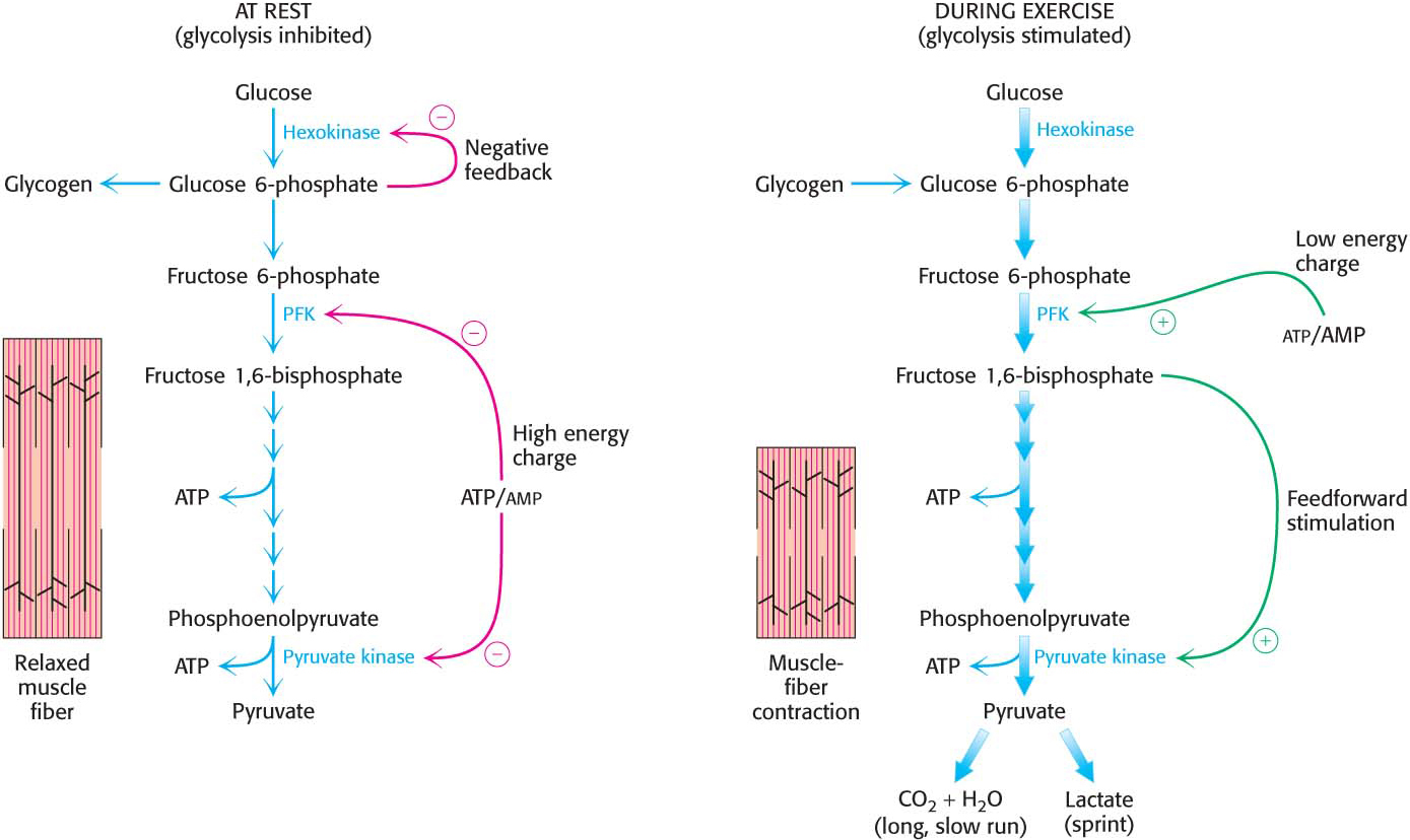
Glycolysis in skeletal muscle provides ATP primarily to power contraction. Consequently, the primary control of muscle glycolysis is the energy charge of the cell—the ratio of ATP to AMP. Glycolysis is stimulated as the energy charge falls—a signal that the cell needs more ATP. Let us examine how each of the key regulatory enzymes responds to changes in the amounts of ATP and AMP present in the cell.

PhosphofructokinasePhosphofructokinase is the most important control site in the mammalian glycolytic pathway. High levels of ATP allosterically inhibit the enzyme (a 340-kDa tetramer). ATP binds to a specific regulatory site that is distinct from the catalytic site. The binding of ATP lowers the enzyme’s affinity for fructose 6-phosphate. AMP reverses the inhibitory action of ATP. AMP competes with ATP for the binding site but, when bound, does not inhibit the enzyme. Consequently the activity of the enzyme increases when the ATP/AMP ratio is lowered (Figure 16.12). A decrease in pH also inhibits phosphofructokinase activity by augmenting the inhibitory effect of ATP. The pH might fall when fast-twitch muscle is functioning anaerobically, producing excessive quantities of lactic acid. The inhibition of glycolysis, and therefore of lactic acid fermentation, protects the muscle from damage that would result from the accumulation of too much acid.

Figure 16.12: The allosteric regulation of phosphofructokinase. A high level of ATP inhibits the enzyme by decreasing its affinity for fructose 6-phosphate. AMP diminishes the inhibitory effect of ATP.

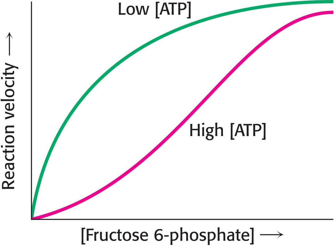
Why does AMP but not ADP stimulate the activity of phosphofructokinase? When ATP is being utilized rapidly, the enzyme adenylate kinase can form ATP from ADP by the following reaction:

Thus, some ATP is salvaged from ADP, and AMP becomes the signal for the low-energy state (problem 16.38).

300

HexokinasePhosphofructokinase is the primary regulatory enzyme in glycolysis, but it is not the only one. Hexokinase, the enzyme catalyzing the first step of glycolysis, is inhibited by its product, glucose 6-phosphate. High concentrations of glucose 6-phosphate signal that the cell no longer requires glucose for energy, and so no more glucose needs to be broken down. The glucose will then be left in the blood. A rise in glucose 6-phosphate concentration is a means by which phosphofructokinase communicates with hexokinase. When phosphofructokinase is inactive, the concentration of fructose 6-phosphate rises. In turn, the level of glucose 6-phosphate rises because it is in equilibrium with fructose 6-phosphate. Hence, the inhibition of phosphofructokinase leads to the inhibition of hexokinase.

Why is phosphofructokinase rather than hexokinase the pacemaker of glycolysis? The reason becomes evident on noting that glucose 6-phosphate is not solely a glycolytic intermediate. In muscle, for example, glucose 6-phosphate can also be converted into glycogen. The first irreversible reaction unique to the glycolytic pathway, the committed step, is the phosphorylation of fructose 6-phosphate to fructose 1,6-bisphosphate. Thus, phosphofructokinase as the primary control site in glycolysis is highly appropriate. In general, the enzyme catalyzing the committed step in a metabolic sequence is the most important control element in the pathway because it regulates flux down the pathway.

Pyruvate kinasePyruvate kinase, the enzyme catalyzing the third irreversible step in glycolysis, controls the efflux from this pathway. This final step yields ATP and pyruvate, a central metabolic intermediate that can be oxidized further or used as a building block. ATP allosterically inhibits pyruvate kinase to decrease the rate of glycolysis when the energy charge of the cell is high. When the pace of glycolysis increases, fructose 1,6-bisphosphate, the product of the preceding irreversible step in glycolysis, activates the kinase to enable it to keep pace with the oncoming high flux of intermediates. A summary of the regulation of glycolysis in resting and active muscle is shown in Figure 16.13.

Figure 16.13: The regulation of glycolysis in muscle. At rest (left), glycolysis is not very active (thin arrows). The high concentration of ATP inhibits phosphofructokinase (PFK) and pyruvate kinase, while glucose 6-phosphate inhibits hexokinase. Glucose 6-phosphate is converted into glycogen (Chapter 25). During exercise (right), the decrease in the ATP/AMP ratio resulting from muscle contraction activates phosphofructokinase and hence glycolysis. The flux down the pathway is increased, as represented by the thick arrows.

The Regulation of Glycolysis in the Liver Corresponds to the Biochemical Versatility of the Liver

The liver has a greater diversity of biochemical functions than muscle. Significantly, the liver maintains blood-glucose concentration: it stores glucose as glycogen when glucose is plentiful, and it releases glucose when supplies are low. It also uses glucose to generate reducing power for biosynthesis (Chapter 26) as well as to synthesize a host of building blocks for other biomolecules. So, although the liver has many of the regulatory features of muscle glycolysis, the regulation of glycolysis in the liver is more complex.

PhosphofructokinaseLiver phosphofructokinase can be regulated by ATP as in muscle, but such regulation is not as important since the liver does not experience the sudden ATP needs that a contracting muscle does. Likewise, low pH is not a metabolic signal for the liver enzyme, because lactate is not normally produced in the liver. Indeed, as we will see, lactate is converted into glucose in the liver.

Glycolysis in the liver furnishes carbon skeletons for biosyntheses, and so a signal indicating whether building blocks are abundant or scarce should also regulate phosphofructokinase. In the liver, phosphofructokinase is inhibited by citrate, an early intermediate in the citric acid cycle (Chapter 19). A high level of citrate in the cytoplasm means that biosynthetic precursors are abundant, and so there is no need to degrade additional glucose for this purpose. In this way, citrate enhances the inhibitory effect of ATP on phosphofructokinase.

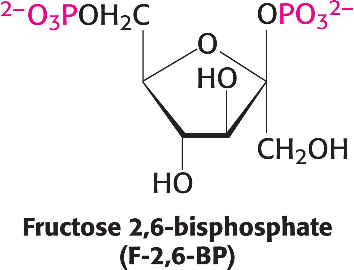
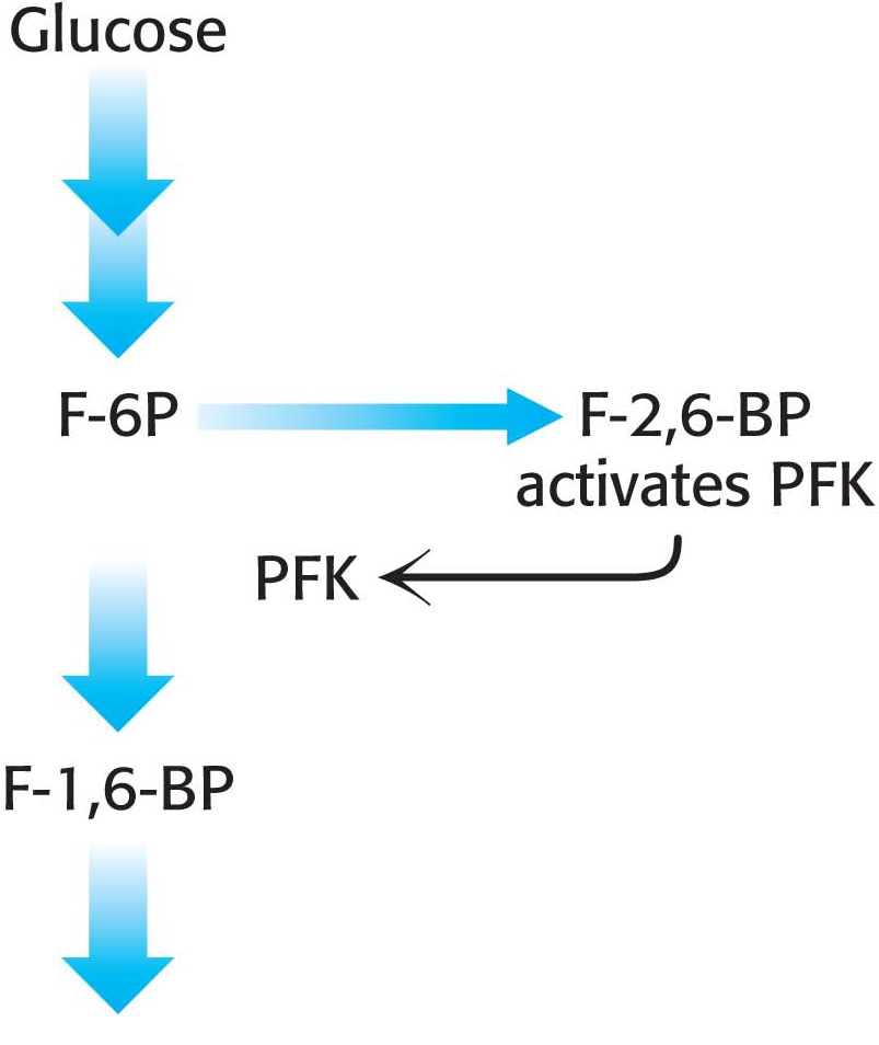
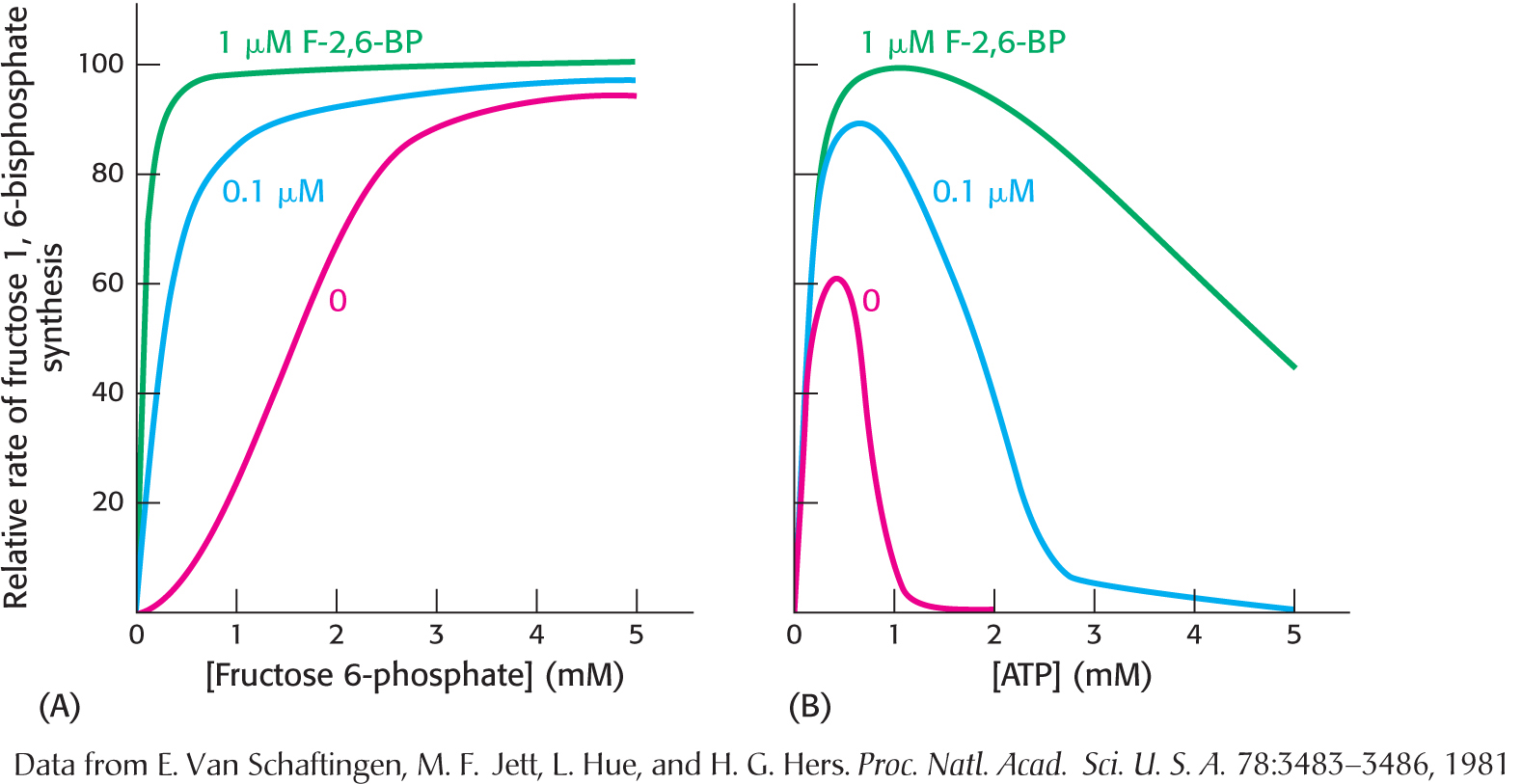
The key means by which glycolysis in the liver responds to changes in blood glucose is through the signal molecule fructose 2,6-bisphosphate (F-2,6-BP), a potent activator of phosphofructokinase. After a meal rich in carbohydrates, the concentration of glucose in the blood rises. In the liver, the concentration of fructose 6-phosphate rises when blood-glucose concentration is high because of the action of hexokinase and phosphoglucose isomerase, and the abundance of fructose 6-phosphate accelerates the synthesis of F-2,6-BP (Figure 16.14). Hence an abundance of fructose 6-phosphate leads to a higher concentration of F-2,6-BP. Fructose 2,6-biphosphate stimulates glycolysis by increasing phosphofructokinase’s affinity for fructose 6-phosphate and diminishing the inhibitory effect of ATP (Figure 16.15). Glycolysis is thus accelerated when glucose is abundant. Such a process is called feedforward stimulation. We will examine the synthesis and degradation of this regulatory molecule after we have considered gluconeogenesis (Chapter 17).

Figure 16.14: The regulation of phosphofructokinase by fructose 2,6-bisphosphate. High concentrations of fructose 6-phosphate (F-6P) activate the enzyme phosphofructokinase (PFK) through an intermediary, fructose 2,6-bisphosphate (F-2,6-BP).
Figure 16.15: The activation of phosphofructokinase by fructose 2,6-bisphosphate. (A) The sigmoidal dependence of velocity on substrate concentration becomes hyperbolic in the presence of 1 mM fructose 2,6-bisphosphate, indicating that more of the enzyme is active at lower substrate concentrations in the presence of fructose 2,6-bisphosphate. (B) ATP, acting as a substrate, initially stimulates the reaction. As the concentration of ATP increases, it acts as an allosteric inhibitor. The inhibitory effect of ATP is reduced by fructose 2,6-bisphosphate, which renders the enzyme less sensitive to ATP inhibition.

301

302

Hexokinase and glucokinaseIn the liver as well as in muscle, hexokinase is a regulatory enzyme. The hexokinase reaction is controlled in the liver as in muscle. However, the enzyme primarily responsible for phosphorylating glucose in the liver is not hexokinase, but glucokinase (hexokinase IV), an isozyme of hexokinase. Isozymes, or isoenzymes, are enzymes encoded by different genes with different amino acid sequences, yet they catalyze the same reaction. Isozymes usually differ in kinetic or regulatory properties. Glucokinase phosphorylates glucose only when glucose is abundant, as would be the case after a meal. The reason is that glucokinase’s KM for glucose is about 50-fold higher than that of hexokinase, which means that glucose 6-phosphate is formed only when glucose is abundant. Furthermore, glucokinase is not inhibited by its product, glucose 6-phosphate, as hexokinase is. The high KM of glucokinase for glucose in the liver gives the brain and muscles first call for glucose when its supply is limited, and it ensures that glucose will not be wasted when it is abundant. The role of glucokinase is to provide glucose 6-phosphate for the synthesis of glycogen and for the formation of fatty acids. Drugs that activate liver glucokinase are being evaluated as a treatment for type 2 or insulin-insensitive diabetes.

Glucokinase is also present in the ß cells of the pancreas, where the increased formation of glucose 6-phosphate by glucokinase when the blood-glucose concentration is high leads to the secretion of the hormone insulin. Insulin signals the need to remove glucose from the blood for storage as glycogen or conversion into fat.

Figure 16.16: The control of the catalytic activity of pyruvate kinase. Pyruvate kinase is regulated by allosteric effectors and covalent modification. Fructose 1,6-bisphosphate allosterically stimulates the enzyme, while ATP and alanine (in liver) are allosteric inhibitors. Glucagon, secreted in response to low blood glucose, promotes phosphorylation and inhibition of the enzyme. When blood-glucose levels are adequate, the enzyme is dephosphorylated and activated.

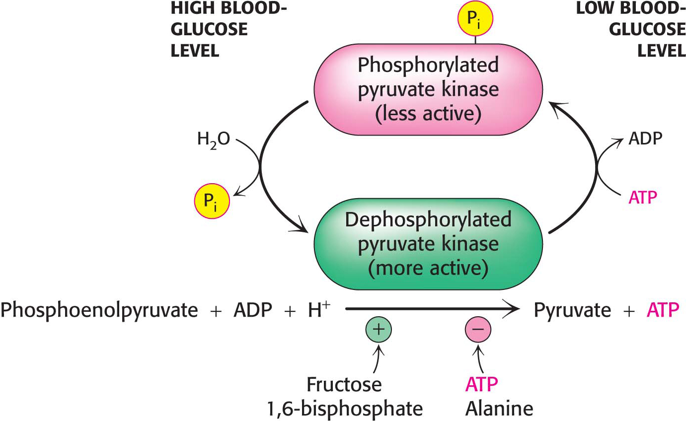
Pyruvate kinaseSeveral isozymic forms of pyruvate kinase (a tetramer of 57-kDa subunits) encoded by different genes are present in mammals: the L form predominates in liver, and the M form in muscle and brain. The L and M forms of pyruvate kinase have many properties in common. Indeed, the liver enzyme behaves much as the muscle enzyme does in regard to allosteric regulation, except that the liver enzyme is also inhibited by alanine (synthesized in one step from pyruvate), a signal that building blocks are available. Moreover, the isozymic forms differ in their susceptibility to phosphorylation. The catalytic properties of the L form—but not of the M form—are controlled by reversible phosphorylation (Figure 16.16). When the blood-glucose concentration is low, the glucagon-triggered cyclic AMP cascade leads to the phosphorylation of pyruvate kinase, which diminishes its activity. This hormone-triggered phosphorylation prevents the liver from consuming glucose when it is more urgently needed by brain and muscle. We see here a clear-cut example of how isoenzymes contribute to the metabolic diversity of different organs. We will return to the control of glycolysis after considering gluconeogenesis (Chapter 17).

303

A Family of Transporters Enables Glucose to Enter and Leave Animal Cells

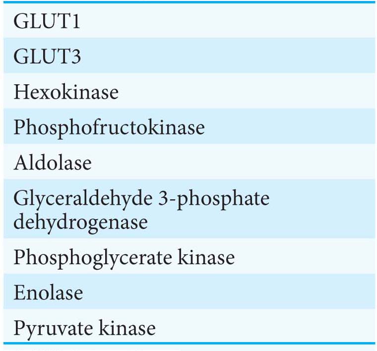
Several glucose transporters mediate the thermodynamically downhill movement of glucose across the plasma membranes of animal cells. Each member of this protein family, named GLUT1 to GLUT5, consists of a single polypeptide chain of about 500 amino acids (Table 16.3).

Table 16.3 Family of glucose transporters

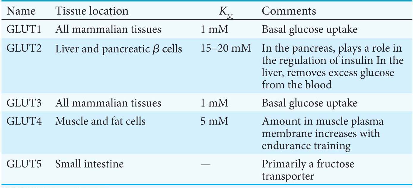
The members of this family have distinctive roles:

  1. GLUT1 and GLUT3, present in nearly all mammalian cells, are responsible for transporting glucose into the cell under normal conditions. Like enzymes, transporters have KM values, except that, for transporters, KM is the concentration of the chemical transported that yields one-half maximal transport velocity. The KM value for glucose for GLUT1 and GLUT3 is about 1 mM, significantly less than the normal serum-glucose concentration, which typically ranges from 4 mM to 8 mM. Hence, GLUT1 and GLUT3 continuously transport glucose into cells at an essentially constant rate.

  2. GLUT2, present in liver and pancreatic ß cells, is distinctive in having a very high KM value for glucose (15–20 mM). Hence, glucose enters these tissues at a biologically significant rate only when there is much glucose in the blood. The pancreas can thereby sense the glucose level and adjust the rate of insulin secretion accordingly. Insulin signals the need to remove glucose from the blood for storage as glycogen or conversion into fat (Chapters 25 and 28). The high KM value of GLUT2 also ensures that glucose rapidly enters liver cells only in times of plenty.

  3. GLUT4, which has a KM value of 5 mM, transports glucose into muscle and fat cells. The number of GLUT4 transporters in the plasma membrane increases rapidly in the presence of insulin, which signals the presence of glucose in the blood. Hence, insulin promotes the uptake of glucose by muscle and fat. Endurance exercise training also increases the amount of GLUT4 present in muscle membranes by a means independent of insulin.

  4. GLUT5, present in the small intestine, functions primarily as a fructose transporter.

304

!clinic! CLINICAL INSIGHT: Aerobic Glycolysis Is a Property of Rapidly Growing Cells

Tumors have been known for decades to display enhanced rates of glucose uptake and glycolysis. Indeed, rapidly growing tumor cells will metabolize glucose to lactate even in the presence of oxygen, a process called aerobic glycolysis or the “Warburg effect,” after Otto Warburg, the biochemist who first noted this characteristic of cancer cells in the 1920s. In fact, tumors with a high glucose uptake are particularly aggressive, and the cancer is likely to have a poor prognosis. A nonmetabolizable glucose analog, 2-18F-2-D-deoxyglucose, detectable by a combination of positron emission tomography (PET) and computer-aided tomography (CAT), easily visualizes tumors (Figure 16.17).

Figure 16.17: Tumors can be visualized with 2-18F-2-D-deoxyglucose (FDG) and positron emission tomography. (A) A nonmetabolizable glucose analog (FDG) infused into a patient and detected by a combination of positron emission and computer-aided tomography reveals the presence of a malignant tumor (T). (B) After 4 weeks of treatment with a tyrosine kinase inhibitor, the tumor shows no uptake of FDG, indicating decreased metabolism. Excess FDG, which is excreted in the urine, also visualizes the kidney (K) and bladder (B).

What selective advantage does aerobic glycolysis offer the tumor over the energetically more efficient oxidative phosphorylation? Research is being actively pursued to answer the question, but we can speculate on the benefits. First, aerobic glycolysis generates lactic acid that is then secreted. Acidification of the tumor environment has been shown to facilitate tumor invasion and inhibit the immune system from attacking the tumor. However, even leukemia cells perform aerobic glycolysis, and leukemia is not an invasive cancer. Second, and perhaps more importantly, the increased uptake of glucose and formation of glucose 6-phosphate provides substrates for another metabolic pathway, the pentose phosphate pathway (Chapter 20), that generates biosynthetic reducing power. Moreover, the pentose phosphate pathway, in cooperation with glycolysis, produces precursors for biomolecules necessary for growth, such as nucleotides. Finally, cancer cells grow more rapidly than the blood vessels that nourish them; thus, as solid tumors grow, the oxygen concentration in their environment falls. In other words, they begin to experience hypoxia, a deficiency of oxygen. The use of aerobic glycolysis reduces the dependence of cell growth on oxygen. Not all of the precursor needs are met by enhanced glucose metabolism. Cancer cells also require glutamine, which is channeled into the mitochondria to replenish citric acid cycle components used for biosynthesis.

305

What biochemical alterations facilitate the switch to aerobic glycolysis? Again, the answers are not complete, but changes in gene expression of isozymic forms of two glycolytic enzymes may be crucial. Tumor cells express an isozyme of hexokinase that binds to mitochondria. There, the enzyme has ready access to any ATP generated by oxidative phosphorylation and is not susceptible to feedback inhibition by its product, glucose 6-phosphate. More importantly, an embryonic isozyme of pyruvate kinase, pyruvate kinase M, is also expressed. Remarkably, this isozyme has a lower catalytic rate than normal pyruvate kinase and creates a bottleneck, allowing the use of glycolytic intermediates for biosynthetic processes required for cell proliferation. The need for biosynthetic precursors is greater than the need for ATP, suggesting that even glycolysis at a reduced rate produces sufficient ATP to allow cell proliferation. Although originally discovered in cancer cells, the Warburg effect is also observed in noncancerous rapidly dividing cells.

Table 16.4 Proteins in glucose metabolism encoded by genes regulated by hypoxia-inducible factor

!clinic! CLINICAL INSIGHT: Cancer and Exercise Training Affect Glycolysis in a Similar Fashion

The hypoxia that some tumors experience with rapid growth activates a transcription factor, hypoxia-inducible transcription factor (HIF-1). HIF-1 increases the expression of most glycolytic enzymes and the glucose transporters GLUT1 and GLUT3 (Table 16.4). These adaptations by the cancer cells enable a tumor to survive until blood vessels can grow. HIF-1 also increases the expression of signal molecules, such as vascular endothelial growth factor (VEGF), that facilitate the growth of blood vessels that will provide nutrients to the cells (Figure 16.18). Without new blood vessels, a tumor would cease to grow and either die or remain harmlessly small. Efforts are underway to develop drugs that inhibit the growth of blood vessels in tumors.

Figure 16.18: The alteration of gene expression in tumors because of hypoxia. The hypoxic conditions inside a tumor mass lead to the activation of the hypoxia-inducible transcription factor (HIF-1), which induces metabolic adaptation (an increase in glycolytic enzymes) and activates angiogenic factors that stimulate the growth of new blood vessels. [Information from C. V. Dang and G. L. Semenza, Trends Biochem. Sci. 24:68–72, 1999.]

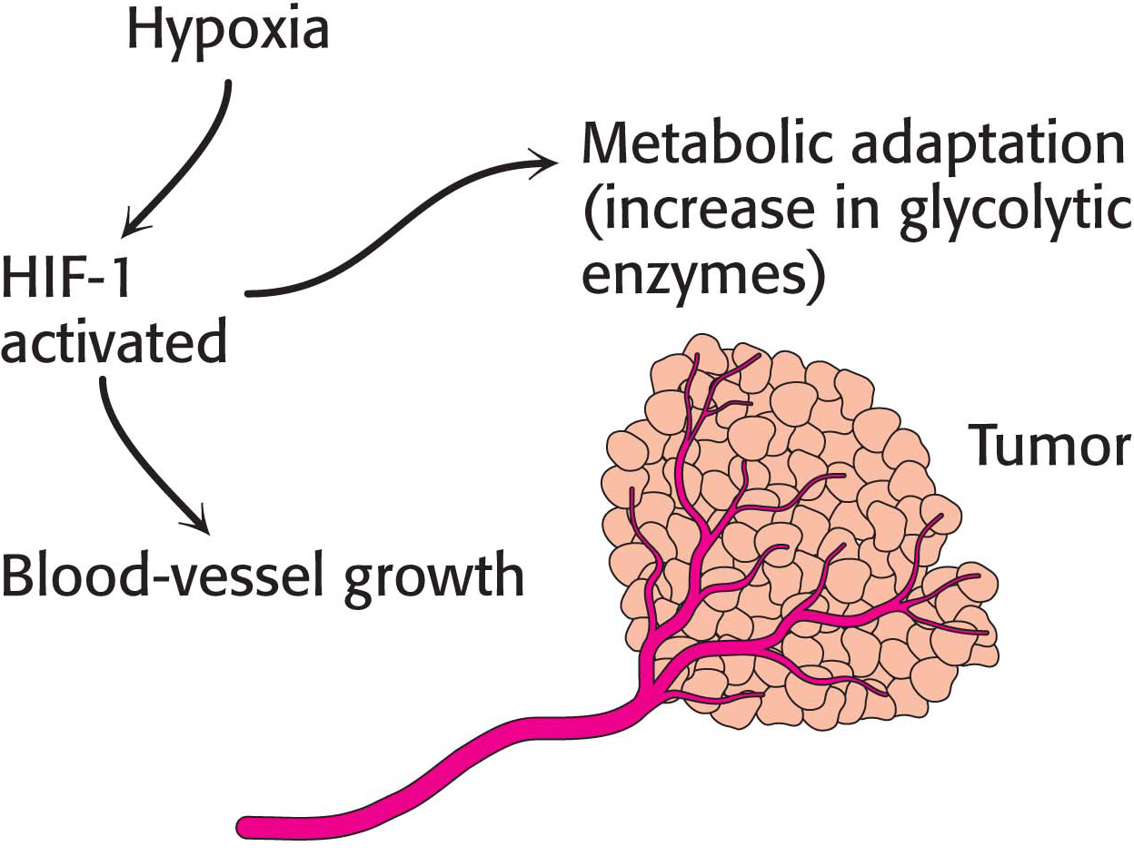
Interestingly, anaerobic exercise training activates HIF-1, producing the same effects as those seen in the tumor—enhanced ability to generate ATP anaerobically and a stimulation of blood-vessel growth. These biochemical effects account for the improved athletic performance that results from training and demonstrate how behavior can affect biochemistry. Other signals from sustained muscle contraction trigger muscle mitochondrial biogenesis, allowing aerobic energy generation and forestalling the need to resort to aerobic glycolysis.

306