20.3 The Respiratory Chain Consists of Proton Pumps and a Physical Link to the Citric Acid Cycle

✓ 2 Explain the benefits of having the electron-transport chain located in a membrane.

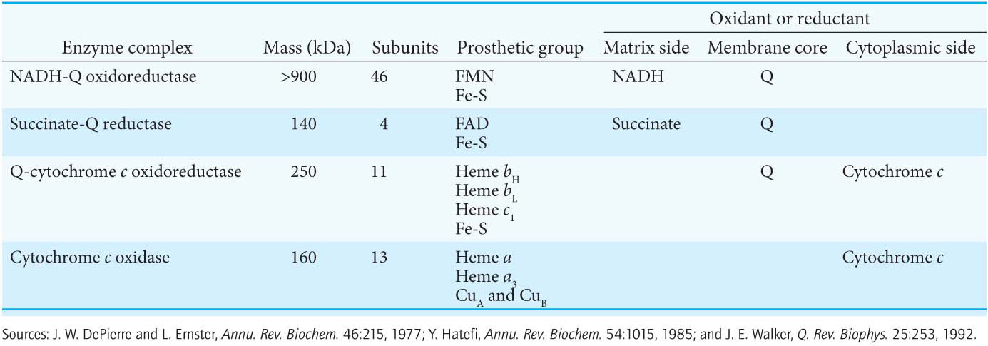
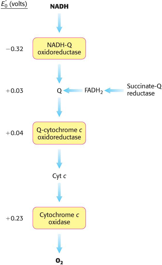
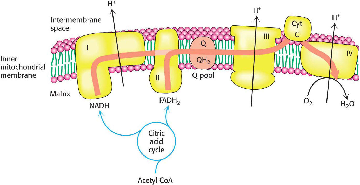
How are the various electron carriers arranged to yield an exergonic reaction that ultimately results in the generation of a proton gradient? As discussed previously (Figure 20.6), electrons are transferred from NADH to O2 through a chain of three protein complexes (Table 20.2 and Figure 20.10). Electron flow within these transmembrane complexes leads to the transport of protons across the inner mitochondrial membrane. A fourth protein complex, succinate-Q reductase, in contrast with the other complexes, does not pump protons.

Table 20.2 Components of the mitochondrial electron-transport chain
Figure 20.10: The components of the electron-transport chain are arranged in complexes. Notice that the electron affinity of the components increases as electrons move down the chain. The complexes shown in yellow boxes are proton pumps. Cyt c stands for cytochrome c.

The High+-Potential Electrons of NADH Enter the Respiratory Chain at NADH-Q Oxidoreductase

The electrons of NADH enter the chain at NADH-Q oxidoreductase (also called Complex I and NADH dehydrogenase), an enormous enzyme (>900 kDa) consisting of approximately 46 polypeptide chains and two types of prosthetic groups: FMN and iron–sulfur clusters. This proton pump is L-shaped, with a hydrophobic horizontal arm lying in the membrane and a hydrophilic vertical arm that projects into the matrix. The electrons flow from NADH to FMN and then through a series of seven iron–sulfur clusters to Q. Note that all of the redox reactions take place in the extramembranous part of NADH-Q oxidoreductase. Although the precise stoichiometry of the reaction catalyzed by this enzyme is not completely worked out, it appears to be

372

Recent structural studies have suggested how Complex I acts as a proton pump. What are the structural elements required for proton pumping? The membrane-embedded part of the complex has four proton half-channels consisting, in part, of vertical helices. One set of half-channels is exposed to the matrix and the other to the intermembrane space (Figure 20.11). The vertical helices are linked on the matrix side by a long horizontal helix (HL) that connects the matrix half-channels, while the intermembrane space half-channels are joined by a series of β-hairpin-helix connecting elements (βH). An enclosed Q chamber, the site where Q accepts electrons from NADH, exists near the junction of the hydrophilic portion and the membrane-embedded portion. Finally, a hydrophilic funnel connects the Q chamber to a water-lined channel, into which the half-channels open, that extends the entire length of the membrane-embedded portion.

NUTRITION FACT

Coenzyme Q is frequently marketed as a dietary supplement, often called CoQ 10. Its proponents assert that it boosts energy, enhances the immune system, and acts as an antioxidant. Clearly, people who lack the ability to synthesize CoQ and consequently suffer from neuromuscular disorders due to defective mitochondria benefit from CoQ supplementation. Moreover, some evidence is beginning to accumulate that shows that CoQ supplementation may benefit people undergoing severe physiological stress—for instance, from heart disease or cancer. However, no studies have established that CoQ supplementation will help a well-nourished person in any activity. In fact, because of its antioxidant effects, CoQ supplementation may actually negate the beneficial effects of exercise (problem 16).

Figure 20.11: Coupled electron–proton transfer reactions through NADH-Q oxidoreductase. Electrons flow in Complex I from NADH through FMN and a series of iron–sulfur clusters to ubiquinone (Q), forming Q2−. The charges on Q2− are electrostatically transmitted to hydrophilic amino acid residues (shown as red and blue balls) that power the movement of HL and bH components. This movement changes the conformation of the transmembrane helices and results in the transport of four protons out of the mitochondrial matrix.

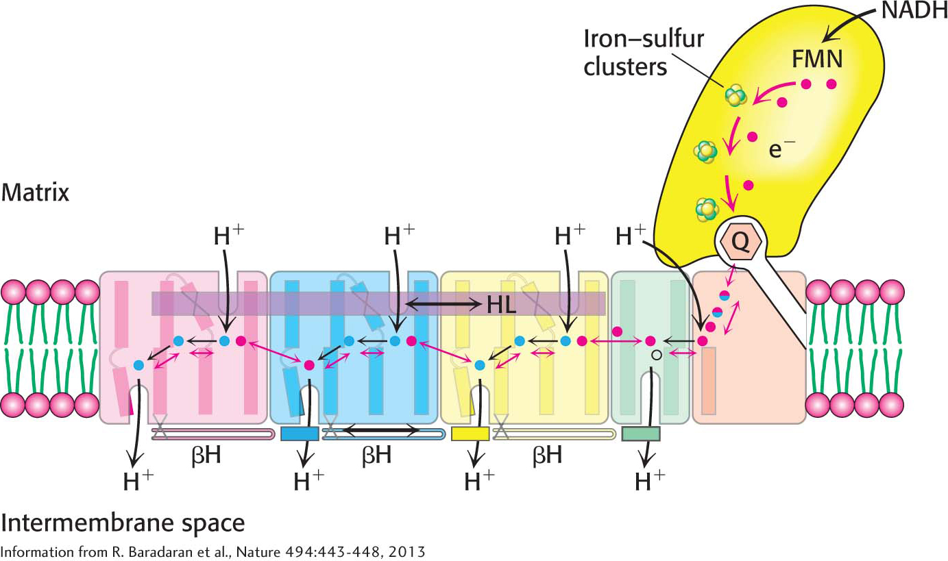
How do these structural elements cooperate to pump protons out of the matrix? When Q accepts two electrons from NADH, generating Q2−,the negative charges on Q2− interact electrostatically with negatively charged amino acid residues in the membrane-embedded arm, causing conformational changes in the long horizontal helix and the bH elements. These changes in turn alter the structures of the connected vertical helices that change the pKa of amino acids, allowing protons from the matrix to first bind to the amino acids, then dissociate into the water-lined channel and finally enter the intermembrane space. Thus, the flow of two electrons from NADH to coenzyme Q through NADH-Q oxidoreductase leads to the pumping of four hydrogen ions out of the matrix of the mitochondrion. Q2− subsequently takes up two protons from the matrix as it is reduced to QH2. The removal of these protons from the matrix contributes to the formation of the proton-motive force. The QH2 subsequently leaves the enzyme for the Q pool, allowing another reaction cycle to occur.

373

Ubiquinol Is the Entry Point for Electrons from FADH2 of Flavoproteins

Recall that FADH2 is formed in the citric acid cycle in the oxidation of succinate to fumarate by succinate dehydrogenase. This enzyme is part of the succinate-Q reductase complex (Complex II), an integral membrane protein of the inner mitochondrial membrane. The electron carriers in this complex are FAD, iron–sulfur proteins, and Q. FADH2 does not leave the complex. Rather, its electrons are transferred to Fe-S centers and then to Q for entry into the electron-transport chain. The succinate-Q reductase complex, in contrast with NADH-Q oxidoreductase, does not transport protons. Consequently, less ATP is formed from the oxidation of FADH2 than from NADH.

Electrons Flow from Ubiquinol to Cytochrome c Through Q-Cytochrome c Oxidoreductase

The second of the three proton pumps in the respiratory chain is Q-cytochrome c oxidoreductase (also known as Complex III and cytochrome c reductase). The function of Q-cytochrome c oxidoreductase is to catalyze the transfer of electrons from QH2 produced by NADH-Q oxidoreductase and the succinate-Q reductase complex to oxidized cytochrome c (Cyt c), a water-soluble protein, and concomitantly pump protons out of the mitochondrial matrix. The flow of a pair of electrons through this complex leads to the effective net transport of 2 H+ to the intermembrane space, half the yield obtained with NADH-Q oxidoreductase because of a smaller thermodynamic driving force.

Q-cytochrome c oxidoreductase, a complex protein composed of 22 subunits, contains a total of three hemes, which themselves are contained within two cytochrome subunits: two hemes, termed heme bL (L for low affinity) and heme bH (H for high affinity) within cytochrome b, and one heme within cytochrome c1. Because of these groups, this enzyme is also known as cytochrome bc1. In addition to the hemes, the enzyme also contains an iron–sulfur protein with a 2Fe-2S center. This center, termed the Rieske center, is unusual because one of the iron ions is coordinated by two histidine residues rather than two cysteine residues.

374

The Q Cycle Funnels Electrons from a Two-Electron Carrier to a One-Electron Carrier and Pumps Protons

QH2 passes two electrons to Q-cytochrome c oxidoreductase, but the acceptor of electrons in this complex, cytochrome c, can accept only one electron. How does the switch from the two-electron carrier ubiquinol to the one-electron carrier cytochrome c take place? The mechanism for the coupling of electron transfer from Q to cytochrome c to transmembrane proton transport is known as the Q cycle (Figure 20.12).

Figure 20.12: The Q cycle. In the first half of the cycle, two electrons of a bound QH2 are transferred, one to cytochrome c and the other, after passing through cytochrome b, to a bound Q in a second binding site to form the semiquinone radical anion Q•. The newly formed Q dissociates and enters the Q pool. In the second half of the cycle, a second QH2 also gives up its electrons, one to a second molecule of cytochrome c and the other to reduce Q•to QH2. This second electron transfer results in the uptake of two protons from the matrix. The path of electron transfer is shown in red.

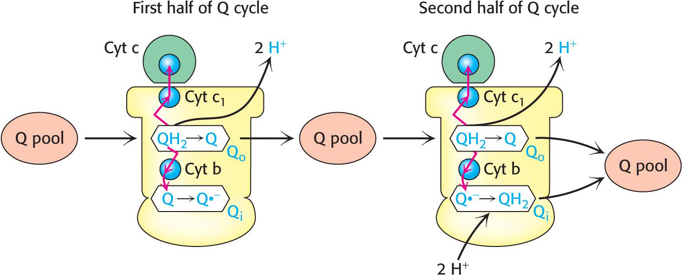
The cycle begins when two QH2 molecules bind to the complex consecutively, each giving up two electrons and two H+. These protons are released to the intermembrane space. QH2 binds to the first Q binding site (Qo), and the two electrons travel through the complex to different destinations. One electron flows, first, to the Rieske 2Fe-2S cluster; then, to cytochrome c1; and, finally, to a molecule of oxidized cytochrome c, converting it into its reduced form. The reduced cytochrome c molecule is free to move away from the enzyme to the final complex of the respiratory chain.

The second electron passes through two heme groups of cytochrome b to an oxidized ubiquinone in a second Q binding site (Qi). The Q in the second binding site is reduced to a semiquinone radical anion (Q?) by the electron from the first QH2. The now fully oxidized Q leaves the first Q site, free to reenter the Q pool.

A second molecule of QH2 binds to Q-cytochrome c oxidoreductase and reacts in the same way as the first. One of the electrons is transferred to cytochrome c. The second electron passes through the two heme groups of cytochrome b to the partly reduced ubiquinone bound in the second binding site. On the addition of the electron from the second QH2 molecule, this quinone radical anion takes up two protons from the matrix side to form QH2. The removal of these two protons from the matrix contributes to the formation of the proton gradient. This complex set of reactions can be summarized as follows: four protons are released into the intermembrane space, and two protons are removed from the mitochondrial matrix.

In one Q cycle, two QH2 molecules are oxidized to form two Q molecules, and then one Q molecule is reduced to QH2. The problem of how to efficiently funnel electrons from a two-electron carrier (QH2) to a one-electron carrier (cytochrome c) is solved by the Q cycle. The cytochrome b component of the reductase is in essence a recycling device that enables both electrons of QH2 to be used effectively.

375

Cytochrome c Oxidase Catalyzes the Reduction of Molecular Oxygen to Water

The last of the three proton-pumping assemblies of the respiratory chain is cytochrome c oxidase (Complex IV). Cytochrome c oxidase catalyzes the transfer of electrons from the reduced form of cytochrome c to molecular oxygen, the final acceptor:

!quickquiz! QUICK QUIZ 1

Why are the electrons carried by FADH2 not as energy rich as those carried by NADH? What is the consequence of this difference?

The requirement of oxygen for this reaction is what makes “aerobic” organisms aerobic. Obtaining oxygen for this reaction is the primary reason that human beings must breathe. Four electrons are funneled to O2 to completely reduce it to two molecules of H2O, and, concurrently, protons are pumped from the matrix to the cytoplasmic side of the inner mitochondrial membrane. This reaction is quite thermodynamically favorable. From the reduction potentials in Table 20.1, the standard free-energy change for this reaction is calculated to be ΔG°′ = –231.8 kJ mol–1 (–55.4 kcal mol–1). As much of this free energy as possible must be captured in the form of a proton gradient for subsequent use in ATP synthesis.

Cytochrome c oxidase, which consists of 13 subunits, contains two heme groups (heme a and heme a3) and three copper ions, arranged as two copper centers (CuA and CuB), with CuA containing two copper ions. Four molecules of reduced cytochrome c generated by Q-cytochrome c oxidoreductase bind consecutively to cytochrome c oxidase and transfer an electron to reduce one molecule of O2 to H2O (Figure 20.13). Let us examine the steps required to generate water at the terminus of the electron-transport chain:

Figure 20.13: The cytochrome c oxidase mechanism. The cycle begins and ends with all prosthetic groups in their oxidized forms (shown in blue). Reduced forms are in red. Four cytochrome c molecules donate four electrons, which, in allowing the binding and cleavage of an O2 molecule, also makes possible the import of four H+ from the matrix to form two molecules of H2O, which are released from the enzyme to regenerate the initial state.

376

Figure 20.14: Proton transport by cytochrome c oxidase. Four protons are taken up from the matrix side to reduce one molecule of O2 to two molecules of H2O. These protons are called “chemical protons” because they participate in a clearly defined reaction with O2. Four additional “pumped” protons are transported out of the matrix and released on the cytoplasmic side in the course of the reaction. The pumped protons double the efficiency of free-energy storage in the form of a proton gradient for this final step in the electron-transport chain.
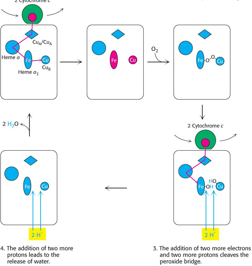
  1. Electrons from two molecules of reduced cytochrome c flow through the oxidation–reduction reactions, one stopping at CuB and the other at heme a3. With both centers in the reduced state, they together can now bind an oxygen molecule.

  2. As molecular oxygen binds, it removes an electron from each of the nearby ions in the active center to form a peroxide (O22−) bridge between them.

  3. Two more molecules of cytochrome c bind and release electrons that travel to the active center. The addition of an electron as well as H+ to each oxygen atom reduces the two ion–oxygen groups to CuB2+—OH and Fe3+—OH.

  4. Reaction with two more H+ ions allows the release of two molecules of H2O and resets the enzyme to its initial, fully oxidized form:

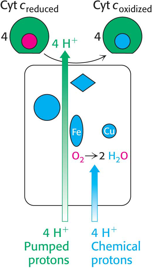
    The four protons in this reaction come exclusively from the matrix. Thus, the consumption of these four protons contributes directly to the proton gradient. Consuming these four protons requires 87.2 kJ mol–1 (19.8 kcal mol–1), an amount substantially less than the free energy released from the reduction of oxygen to water. What is the fate of this missing energy? Remarkably, cytochrome c oxidase uses this energy to pump four additional protons from the matrix to the cytoplasmic side of the membrane in the course of each reaction cycle for a total of eight protons removed from the matrix (Figure 20.14). The details of how these protons are transported through the protein is still under study. Thus, the overall process catalyzed by cytochrome c oxidase is

Figure 20.15 summarizes the flow of electrons from NADH and FADH2 through the respiratory chain. This series of exergonic reactions is coupled to the pumping of protons from the matrix. As we will see in Chapter 21, the energy inherent in the proton gradient will be used to synthesize ATP.

!quickquiz! QUICK QUIZ 2

Amytal is a barbiturate sedative that inhibits electron flow through Complex I. How would the addition of amytal to actively respiring mitochondria affect the relative oxidation–reduction states of the components of the electron-transport chain and the citric acid cycle?

Figure 20.15: The electron-transport chain. High-energy electrons in the form of NADH and FADH2 are generated by the citric acid cycle. These electrons flow through the respiratory chain, which powers proton pumping and results in the reduction of O2.

377

DID YOU KNOW?

In anaerobic respiration in some organisms, chemicals other than oxygen are used as the final electron acceptor in an electron-transport chain. Because none of these electron acceptors are as electropositive as O2, not as much energy is released and, consequently, not as much ATP is generated.

!bio! BIOLOGICAL INSIGHT: 
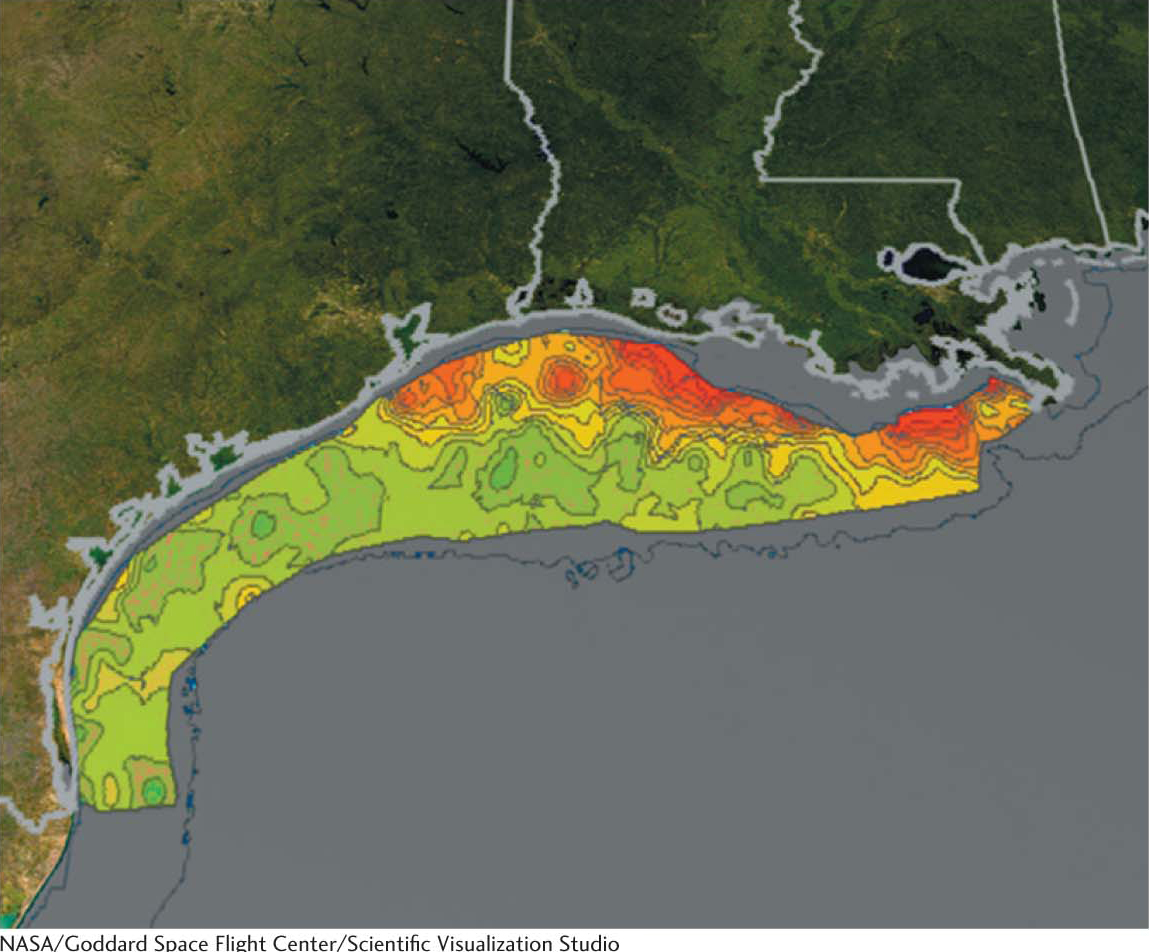
The Dead Zone: Too Much Respiration

Some marine organisms perform so much cellular respiration, and therefore consume so much molecular oxygen, that the oxygen concentration in the water is decreased to a level that is too low to sustain other organisms. One such hypoxic (low levels of oxygen) zone is in the northern Gulf of Mexico, off the coast of Louisiana where the Mississippi River flows into the Gulf (Figure 20.16). The Mississippi is extremely nutrient rich due to agricultural runoff; so plant microorganisms, called phytoplankton, proliferate so robustly that they exceed the amount that can be consumed by other members of the food chain. When the phytoplankton die, they sink to the bottom and are consumed by aerobic bacteria. The aerobic bacteria thrive to such a degree that other bottom-dwelling organisms, such as shrimp and crabs, cannot obtain enough O2 to survive. The term “dead zone” refers to the inability of this area to support fisheries.

Figure 20.16: The gulf of Mexico dead zone. The size of the dead zone in the Gulf of Mexico off Louisiana varies annually but may extend from the Louisiana and Alabama coasts to the westernmost coast of Texas. reds and oranges represent high concentrations of phytoplankton and river sediment.

Toxic Derivatives of Molecular Oxygen Such As Superoxide Radical Are Scavenged by Protective Enzymes

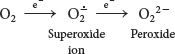
Molecular oxygen is an ideal terminal electron acceptor because its high affinity for electrons provides a large thermodynamic driving force. However, the reduction of O2 can result in dangerous side reactions. The transfer of four electrons leads to safe products (two molecules of H2O), but partial reduction generates hazardous compounds. In particular, the transfer of a single electron to O2 forms superoxide ion, whereas the transfer of two electrons yields peroxide:

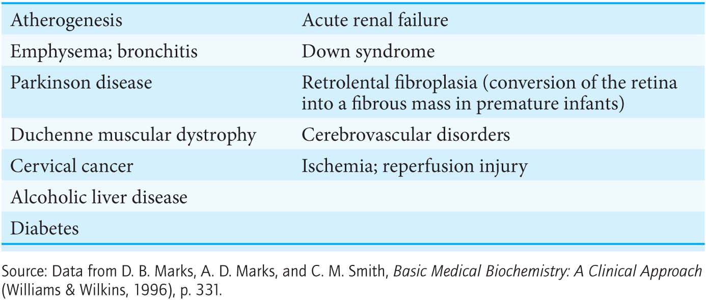
From 2% to 4% of the oxygen molecules consumed by mitochondria are converted into superoxide ion, predominantly at Complexes I and III. Superoxide, peroxide, and species that can be generated from them such as the hydroxyl radical (OH∙) are collectively referred to as reactive oxygen species or ROS. Reactive oxygen species react with essentially all macromolecules in the cell, including proteins, nucleotide bases, and membranes. Oxidative damage caused by ROS has been implicated in the aging process as well as in a growing list of diseases (Table 20.3).

Table 20.3 Pathological and other conditions that may be due to free-radical injury

What are the cellular defense strategies against oxidative damage by ROS? Chief among them is the enzyme superoxide dismutase. This enzyme scavenges superoxide radicals by catalyzing the conversion of two of these radicals into hydrogen peroxide and molecular oxygen:

DID YOU KNOW?

In mammals, the mutation rate for mitochondrial DNA is 10- to 20-fold higher than that for nuclear DNA. This higher rate is believed to be due in large part to the inevitable generation of reactive oxygen species by oxidative phosphorylation in mitochondria.

DID YOU KNOW?

Dismutation is a reaction in which a single reactant is converted into two different products.

378

Figure 20.17: Superoxide dismutase mechanism. The oxidized form of superoxide dismutase (Mox) reacts with one superoxide ion to form O2 and generate the reduced form of the enzyme (Mred). The reduced form then reacts with a second superoxide ion and two protons to form hydrogen peroxide and regenerate the oxidized form of the enzyme.

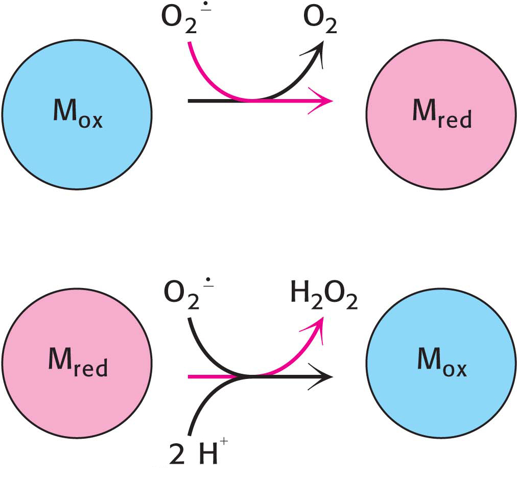
This reaction takes place in two steps. The oxidized form of the enzyme is reduced by superoxide to form oxygen (Figure 20.17). The reduced form of the enzyme then reacts with a second superoxide ion to form peroxide, which takes up two protons along the reaction path to yield hydrogen peroxide.

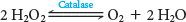
The hydrogen peroxide formed by superoxide dismutase and by other processes is scavenged by catalase, a ubiquitous heme protein that catalyzes the dismutation of hydrogen peroxide into water and molecular oxygen:

Superoxide dismutase and catalase are remarkably efficient, performing their reactions at or near the diffusion-limited rate. Other cellular defenses against oxidative damage include the antioxidant vitamins—vitamins E and C as well as ubiquinol. Because it is lipophilic, vitamin E is especially useful in protecting membranes from lipid peroxidation. Ubiquinol is the only lipid-soluble antioxidant synthesized by human beings.

A long-term benefit of exercise may be to increase the amount of superoxide dismutase in the cell. The elevated aerobic metabolism during exercise causes more ROS to be generated. In response, the cell synthesizes more protective enzymes. The net effect is one of protection, because the increase in superoxide dismutase more effectively protects the cell during periods of rest.

Despite the fact that reactive oxygen species are known hazards, recent evidence suggests that the controlled generation of these molecules may be important components of signal-transduction pathways. For instance, growth factors have been shown to increase ROS as part of their signaling pathway, and ROS regulate channels and transcription factors. ROS have been implicated in the control of cell differentiation, the immune response, autophagy, circadian rhythms as well as other metabolic activities. The dual roles of ROS are an excellent example of the wondrous complexity of the biochemistry of living systems: even potentially harmful substances can be harnessed to play useful roles.