23.1 The Calvin Cycle Synthesizes Hexoses from Carbon Dioxide and Water
✓ 4 Explain the function of the Calvin cycle.
The Calvin cycle takes place in the stroma of chloroplasts, the photosynthetic organelles. In this vital process, carbon dioxide gas is trapped in an organic molecule, 3-phosphoglycerate, which has many biochemical fates.
The Calvin cycle has three stages (Figure 23.1):
The fixation of CO2 by ribulose 1,5-bisphosphate to form two molecules of 3-phosphoglycerate. Recall that 3-phosphoglycerate is an intermediate in glycolysis and gluconeogenesis.
The reduction of 3-phosphoglycerates to form hexose sugars.
The regeneration of ribulose 1,5-bisphosphate so that more CO2 can be fixed (incorporated into an organic molecule).
Figure 23.1: The Calvin cycle. The Calvin cycle consists of three stages. Stage 1 is the fixation of carbon by the carboxylation of ribulose 1,5-bisphosphate. Stage 2 is the reduction of the fixed carbon to begin the synthesis of hexose. Stage 3 is the regeneration of the starting compound, ribulose 1,5-bisphosphate.
Keep in mind that, owing to the stoichiometry of the conversion of six molecules of CO2 into a hexose sugar, to synthesize a glucose molecule or any other hexose sugar, the three stages must take place six times. The regeneration process is a complicated one—hence the dotted arrow and simplification of the process depicted in Figure 23.1; the details will be revealed as we move through the process. For simplicity’s sake, we will consider the reactions one molecule at a time or three molecules at a time.
Carbon Dioxide Reacts with Ribulose 1,5-bisphosphate to Form Two Molecules of 3-Phosphoglycerate
The first stage in the Calvin cycle is the fixation of CO2. This highly exergonic reaction (ΔG°′ = −51.9 kJ mol−1, or −12.4 kcal mol−1) is catalyzed by ribulose 1,5-bisphosphate carboxylase/oxygenase (usually called rubisco), an enzyme located on the stromal surface of the thylakoid membranes of chloroplasts. This reaction is the rate-limiting step in hexose synthesis.
Carbon fixation begins with the conversion of ribulose 1,5-bisphosphate into a highly reactive enediolate intermediate. The CO2 molecule condenses with the enediolate intermediate to form an unstable six-carbon compound, which is rapidly hydrolyzed to two molecules of 3-phosphoglycerate:
Thus, carbon dioxide, a waste product of cellular respiration, is reintroduced to the biochemical world. Plants that use only the Calvin cycle to fix carbon dioxide are called C3 plants, because the three-carbon molecule 3-phosphoglycerate is formed immediately after the rubisco reaction.
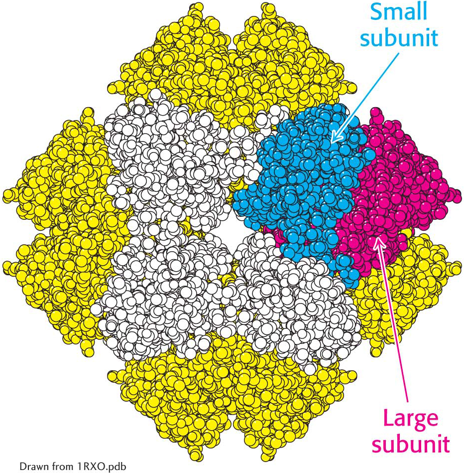
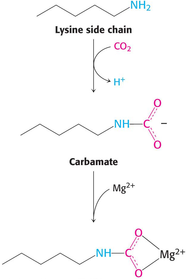
Rubisco in chloroplasts consists of eight large (L, 55-kDa) subunits and eight small (S, 13-kDa) ones (Figure 23.2). Each L chain contains a catalytic site and a regulatory site. The S chains enhance the catalytic activity of the L chains. This enzyme is very abundant in chloroplasts, constituting more than 16% of their total protein. In fact, rubisco is the most abundant enzyme in plants and probably the most abundant protein in the biosphere. Large amounts are required because rubisco is an inefficient enzyme; its maximal catalytic rate is only 3 s−1. Rubisco also requires a bound CO2 for catalytic activity in addition to the substrate CO2. The bound CO2 forms a carbamate with lysine 201 of the large subunit. The carbamate binds to a Mg2+ ion that is required for catalytic activity. We will see the regulatory significance of this requirement later.
Figure 23.2: The structure of rubisco. The enzyme ribulose 1,5-bisphosphate carboxylase/oxygenase (rubisco) comprises eight large subunits (one shown in red and the others in yellow) and eight small subunits (one shown in blue and the others in white). The active sites lie in the large subunits.
In the absence of CO2, rubisco binds to its substrate, ribulose 1,5-bisphosphate, so tightly that its enzyme activity is inhibited. The enzyme rubisco activase uses ATP to induce structural changes in rubisco that allow release of the bound substrate and formation of the required carbamate. The ATP requirement of rubisco activase coordinates rubisco activity with the light reactions.
Hexose Phosphates Are Made from Phosphoglycerate, and Ribulose 1,5-bisphosphate Is Regenerated
In the second stage of the Calvin cycle, the 3-phosphoglycerate products of rubisco are converted into a hexose phosphate. The steps in this conversion (Figure 23.3) are like those of the gluconeogenic pathway, except that glyceraldehyde 3-phosphate dehydrogenase in chloroplasts, which generates glyceraldehyde 3-phosphate (GAP) in the process of hexose formation, is specific for NADPH rather than NADH. The hexose phosphate product of the Calvin cycle exists in three isomeric forms: glucose 1-phosphate, glucose 6-phosphate, and fructose 6-phosphate, together called the hexose monophosphate pool. Recall that these isomers are readily interconvertible. Alternatively, the glyceraldehyde 3-phosphate can be transported to the cytoplasm and metabolized to fructose 6-phosphate and glucose 1-phosphate by using the gluconeogenic pathway and the enzyme phosphoglucomutase. Fructose 6-phosphate and glucose 1-phosphate are used for sucrose synthesis (Figure 23.8). These reactions and the reaction catalyzed by rubisco bring CO2 to the oxidation level of a hexose, converting CO2 into a chemical fuel at the expense of the NADPH and ATP that were generated in the light reactions.
Figure 23.3: Hexose phosphate formation. 3-Phosphoglycerate is converted into fructose 6-phosphate in a pathway parallel to that of gluconeogenesis.
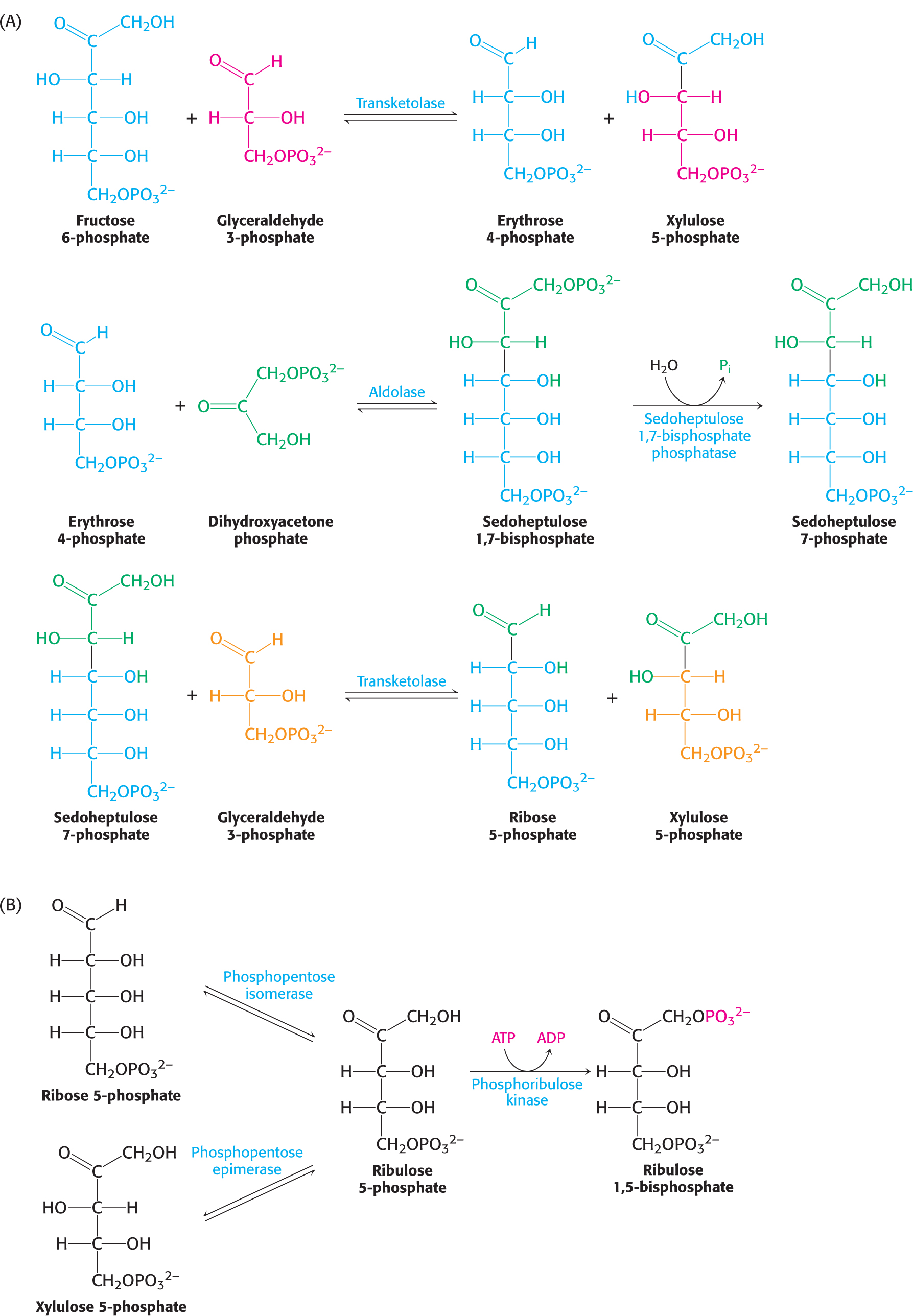
The third and final stage of the Calvin cycle is the regeneration of ribulose 1,5-bisphosphate, the acceptor of CO2 in the first stage. This regeneration is not as simply done as the regeneration of oxaloacetate in the citric acid cycle. The challenge is to construct a five-carbon sugar from a six-carbon member of the hexose monophosphate pool and three-carbon molecules, such as glyceraldehyde 3-phosphate. A transketolase, which we will encounter again in the pentose phosphate pathway, and an aldolase, an enzyme in glycolysis and gluconeogenesis, have major roles in the rearrangement of the carbon atoms. With these enzymes, the construction of the five-carbon sugar proceeds as shown in Figure 23.4. The sum of these reactions is
Figure 23.4: The regeneration of ribulose 1,5-bisphosphate. (A) A transketolase and aldolase generate the five-carbon sugars ribose 5-phosphate and xylulose 5-phosphate from six-carbon and three-carbon sugars. (B) Both ribose 5-phosphate and xylulose 5-phosphate are converted into ribulose 5-phosphate, which is then phosphorylated to complete the regeneration of ribulose 1,5-bisphosphate.
This series of reactions completes the Calvin cycle (Figure 23.5). Figure 23.5 presents the required reactions with the proper stoichiometry to convert three molecules of CO2 into one molecule of dihydroxyacetone phosphate (DHAP). However, two molecules of DHAP are required for the synthesis of a member of the hexose monophosphate pool. Consequently, the cycle as presented must take place twice to yield a hexose monophosphate. The outcome of the Calvin cycle is the generation of a hexose and the regeneration of the starting compound, ribulose 1,5-bisphosphate. In essence, ribulose 1,5-bisphosphate acts catalytically, similarly to oxaloacetate in the citric acid cycle.
Figure 23.5: Stoichiometry of the Calvin cycle. The diagram shows the reactions necessary with the correct stoichiometry to convert three molecules of CO2 into one molecule of dihydroxyacetone phosphate (DHAP). Two molecules of DHAP are subsequently converted into a member of the hexose monophosphate pool, shown here as fructose 6-phosphate. The cycle is not as simple as presented in Figure 23.1; rather, it entails many reactions that lead ultimately to the synthesis of glucose and the regeneration of ribulose 1,5-bisphosphate.
Three Molecules of ATP and Two Molecules of NADPH Are Used to Bring Carbon Dioxide to the Level of a Hexose
What is the energy expenditure for synthesizing a hexose? Six rounds of the Calvin cycle are required, because one carbon atom is reduced in each round. Twelve molecules of ATP are expended in phosphorylating 12 molecules of 3-phosphoglycerate to 1,3-bisphosphoglycerate, and 12 molecules of NADPH are consumed in reducing 12 molecules of 1,3-bisphosphoglycerate to glyceraldehyde 3-phosphate. An additional 6 molecules of ATP are spent in regenerating ribulose 1,5-bisphosphate. The balanced equation for the net reaction of the Calvin cycle is then
Thus, 3 molecules of ATP and 2 molecules of NADPH are consumed in incorporating a single CO2 molecule into a hexose such as glucose or fructose. Biochemically, the synthesis of glucose from CO2 is energetically expensive, but the ultimate energy source—sunlight—is abundant.
Figure 23.6: The eruption of Mount Pinatubo.
!bio! BIOLOGICAL INSIGHT: A Volcanic Eruption Can Affect Photosynthesis Worldwide
On June 12, 1991, Mount Pinatubo, a volcano on the island of Luzon in the Philippines, erupted (Figure 23.6). The eruption, which was the largest on Earth in a hundred years, killed more than 700 people. The eruption also spewed approximately 15 million tons of sulfur dioxide (SO2) into the atmosphere in the form of sulfate aerosol, encircling Earth in a massive aerosol blanket in about 3 weeks. This blanket led to a decrease in total sunlight striking Earth (direct sunlight plus diffuse sunlight) but increased the proportion of diffuse sunlight.
At the same time as the sulfate aerosol was enveloping the planet, the rate of growth of atmospheric CO2 declined sharply. Moreover, an experimental station in the northeastern United States reported that noontime photosynthesis increased by almost 25%. Could these events have been related?
If the increase in photosynthesis observed in the northeastern United States were happening globally, as might have been expected, then more CO2 would have been removed from the atmosphere by rubisco, accounting for the decrease in the rate of growth of CO2. But why did it happen? Although still not firmly established, the diffuse light caused by the sulfate aerosol may have actually increased the amount of photosynthesis taking place. Figure 23.7 shows that the rate of photosynthesis reaches a limiting value when light intensity is increased. Consequently, in bright direct sunlight, the top leaves of a forest may be receiving more light than they can biochemically use. However, in direct sunlight, the lower leaves in the shadows of the upper leaves will be light deprived. Diffuse light, on the other hand, will penetrate more deeply into the forest. More leaves will receive light, more rubisco will be active, and more CO2 will be converted into sugar.
Figure 23.7: Photosynthesis has a maximum rate. The rate of photosynthesis reaches a limiting value when the light-harvesting complexes are saturated with light.
Starch and Sucrose Are the Major Carbohydrate Stores in Plants
What are the fates of the carbon atoms fixed and processed by the enzymes of the Calvin cycle? These molecules are used in a variety of ways, but there are two prominent fates: the synthesis of starch and sucrose, storage forms of carbohydrates. Starch, like its animal counterpart glycogen, is a polymer of glucose residues. Starch comes in two forms: amylose, which consists of glucose molecules joined together by α-1,4 linkages only; and amylopectin, which is branched because of the presence of α-1,6 linkages. Starch is synthesized and stored in chloroplasts.
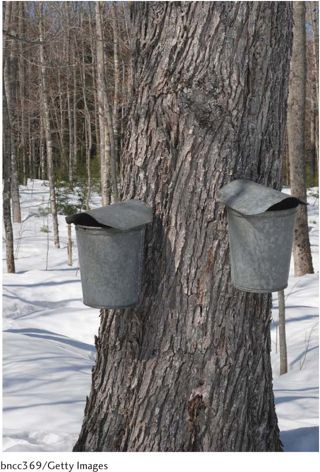
In contrast, sucrose (common table sugar), a disaccharide, is synthesized in the cytoplasm. Plants lack the ability to transport hexose phosphates across the chloroplast membrane, but an abundant triose phosphatephosphate antiporter mediates the transport of triose phosphates from chloroplasts to the cytoplasm in exchange for phosphate. Fructose 6-phosphate is formed from these triose phosphates in the cytoplasm and joins the glucose unit of cytoplasmic UDP-glucose (activated glucose) to form sucrose 6-phosphate (Figure 23.8). Sucrose 6-phosphate is then hydrolyzed by sucrose 6-phosphatase to yield sucrose. Sucrose is transported from the leaves as a component of sap to the nonphotosynthetic parts of the plant, such as the roots, and is stored in many plant cells, as in sugar beets and sugar cane (Figure 23.9).
Figure 23.8: The synthesis of sucrose. Sucrose 6-phosphate is formed by the reaction between fructose 6-phosphate and the activated intermediate uridine diphosphate glucose (UDP-glucose). Sucrose phosphatase subsequently generates free sucrose (not shown).
Figure 23.9: Sucrose is transported through plants as an ingredient of sap. Maple syrup, derived from the sap of the sugar maple by boiling and evaporation, is a solution of approximately 65% sucrose with some glucose and fructose.
!quickquiz! QUICK QUIZ 1
Why is the Calvin cycle crucial to the functioning of all life forms?
!bio! BIOLOGICAL INSIGHT: Why Bread Becomes Stale: The Role of Starch
Bread staling (the process of becoming stale) is a complex process in which all of the components of bread take part—including proteins, lipids, and starch and their complex interactions. Let us consider the role of starch. A granule of starch is normally a strictly organized crystal structure rather than an amorphous form. This order is imparted to the starch granule by hydrogen bonds that form between chains of amylose and amylopectin molecules as they line up next to one another. However, when the starch is heated during baking, the hydrogen bonds of the crystal structure are disrupted, allowing water to be absorbed. The water breaks the crystal structure completely. The starch granules thus become more amorphous, contributing to the taste and texture of the bread crumb (the inside of the loaf of bread).
As the bread cools after it has been removed from the oven (Figure 23.10), amylose molecules separate from the amorphous starch structure and begin to associate with one another; that is, the amylose crystallizes. This crystallization takes place most prominently on the surface of the bread, where it is facilitated by the evaporation of surface water. This crystallization contributes to the crispness of the surface.
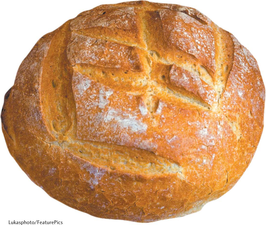
Figure 23.10: Bread staling. Starch crystallization is responsible for the crust of bread.
Meanwhile, inside the bread, crystallization of the amylopectin of the crumb is slower, possibly because of its branched structure, and continues for several days. Water bound to the amylopectin is released as the crystals form, which leads to a deterioration of the texture and taste of the crumb. Water is still present, but no longer in association with the amylopectin molecules. In fact, staling can be reversed at this stage if the bread is reheated to rehydrate the internal amylose and amylopectin crystals. However, if the bread is left uncovered, water will move to the surface, destroying the crispness of the bread and subsequently evaporating. Further evaporation leads to further crystallization, and, eventually, we are left with a loaf of bread more suitable for use as a doorstop.