
33.1 A Nucleic Acid Consists of Bases Linked to a Sugar–Phosphate Backbone
✓ 1 List the components of nucleic acids.
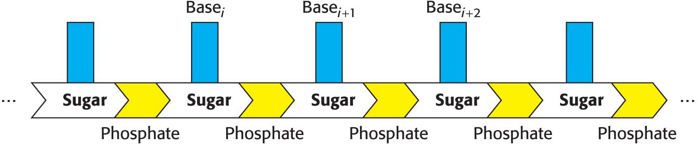
The nucleic acids DNA and RNA are well suited to functioning as the carriers of genetic information by virtue of their structures. These macromolecules are linear polymers built up from similar units connected end to end (Figure 33.2). Each monomer unit within the polymer consists of three components: a sugar, a phosphate, and a base. The sequence of bases uniquely characterizes a nucleic acid and is a form of linear information.
DNA and RNA Differ in the Sugar Component and One of the Bases
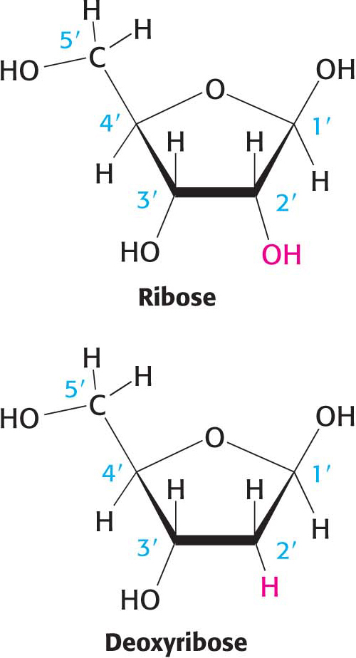
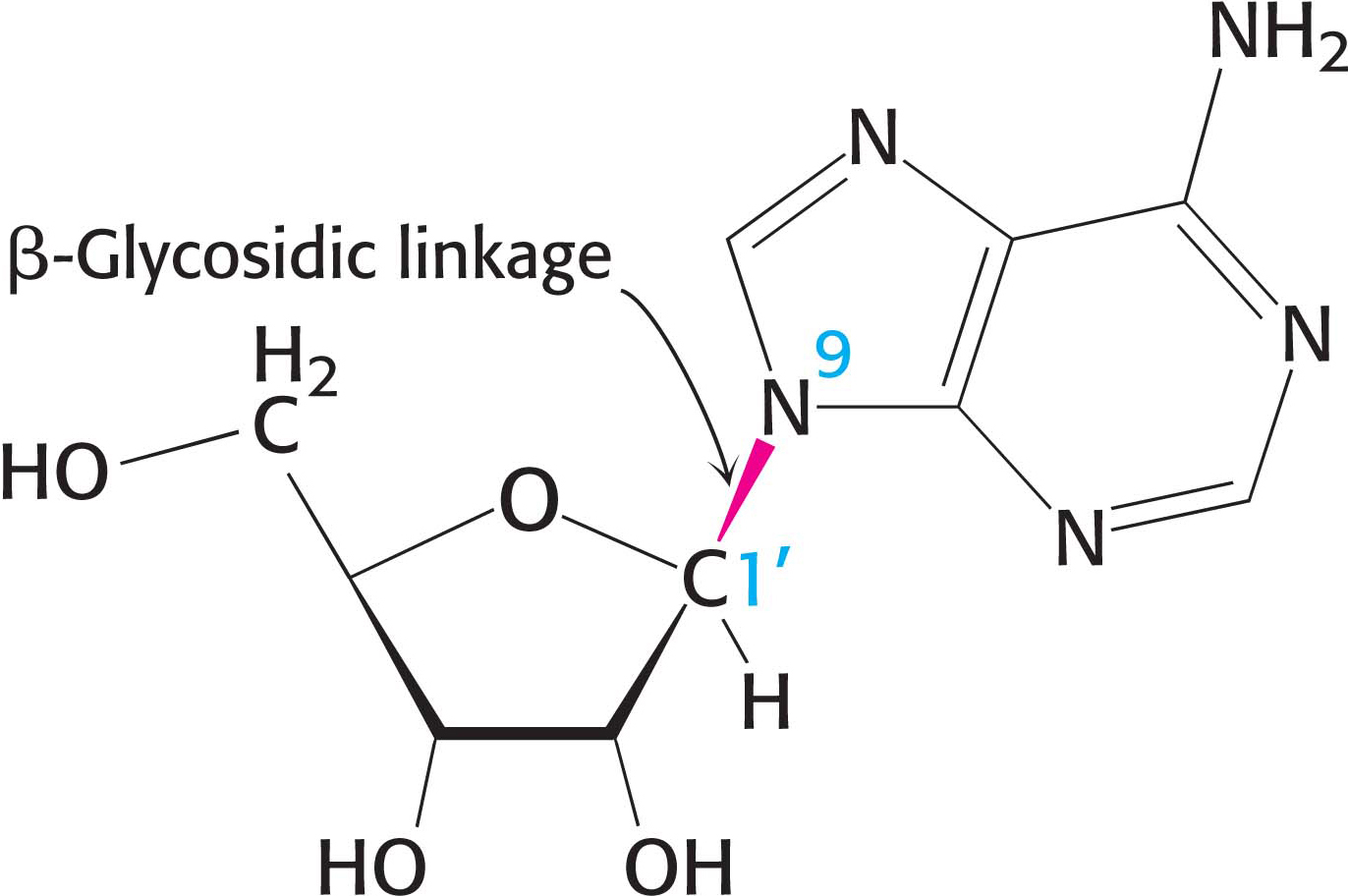
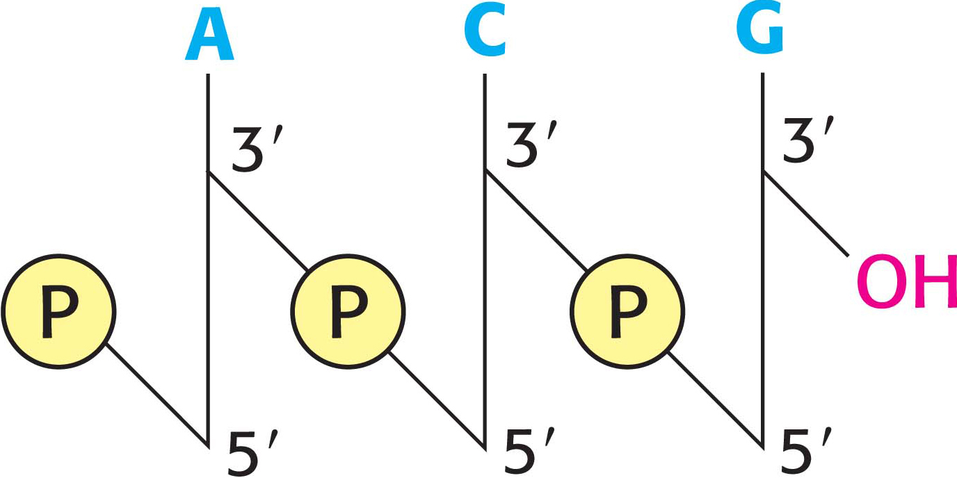
The sugar in deoxyribonucleic acid (DNA) is deoxyribose. The “deoxy” prefix refers to the fact that the 2′-carbon atom of the sugar lacks the oxygen atom that is linked to the 2′-carbon atom of ribose (the sugar in ribonucleic acid, or RNA), as shown in Figure 33.3. The sugars in nucleic acids are linked to one another by phosphodiester bridges. Specifically, the 3′-hydroxyl (3′-OH) group of the sugar component of one nucleotide is bonded to a phosphoryl group, and the phosphoryl group is, in turn, joined to the 5′-hydroxyl group of the adjacent sugar, forming a phosphodiester linkage. The strand of sugars linked by phosphodiester bridges is referred to as the backbone of the nucleic acid (Figure 33.4). The backbone is constant in DNA and RNA, but the bases vary from one monomer to the next. Two of the bases are derivatives of purine—adenine and guanine—
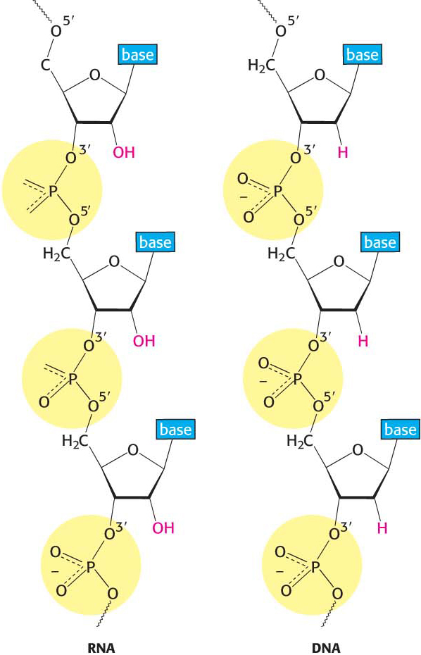
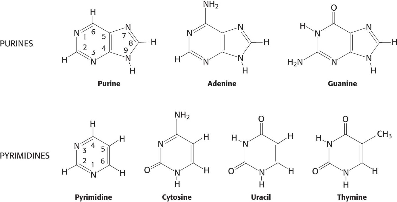
Ribonucleic acid (RNA), like DNA, is a long unbranched polymer consisting of nucleotides joined by 3′ → 5′ phosphodiester linkages (Figure 33.4). The structure of RNA differs from that of DNA in two respects: (1) the sugar units in RNA are riboses rather than deoxyriboses and (2) one of the four major bases in RNA is uracil instead of thymine (Figure 33.5).
Note that each phosphodiester linkage in the backbone of both DNA and RNA has a negative charge. This negative charge repels nucleophilic species such as hydroxide ion, which are capable of hydrolytically cleaving the phosphodiester linkages of the nucleic acid backbone. This resistance is crucial for maintaining the integrity of information stored in nucleic acids. The absence of the 2′-hydroxyl group in DNA further increases its resistance to hydrolysis. In the presence of nucleophilic species, a 2′-hydroxyl group would hydrolyze the phosphodiester linkage and cause a break in the nucleic acid backbone. The greater stability of DNA is one of the reasons for its use rather than RNA as the hereditary material in all modern cells and in many viruses.
Nucleotides Are the Monomeric Units of Nucleic Acids
We considered the synthesis of nucleotides, the building blocks of nucleic acids, in Chapter 32. Let’s review the nomenclature of these crucial biomolecules. A unit consisting of a base bonded to a sugar is referred to as a nucleoside. The four nucleoside units in DNA are called deoxyadenosine, deoxyguanosine, deoxycytidine, and thymidine. Note that thymidine contains deoxyribose; however, by convention, the prefix “deoxy” is not added, because thymine-
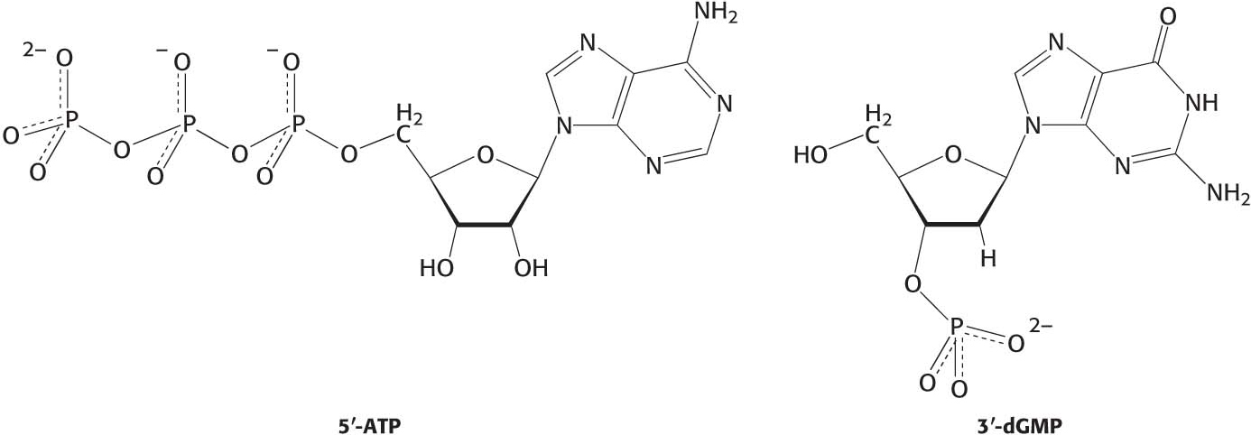
A nucleotide is a nucleoside joined to one or more phosphoryl groups by an ester linkage and is most commonly referred to as a nucleoside with the number of attached phosphoryl groups noted. For instance, a nucleoside monophosphate is a nucleotide. Nucleoside triphosphates are the monomers—
The base name with the suffix “ate” alone is a nucleotide, but with an unknown number of phosphoryl groups attached at an undesignated carbon atom of the sugar. A more precise nomenclature also is commonly used. A compound formed by the attachment of a phosphoryl group to C-
DNA Molecules Are Very Long and Have Directionality
Scientific communication frequently requires the sequence of a nucleic acid—
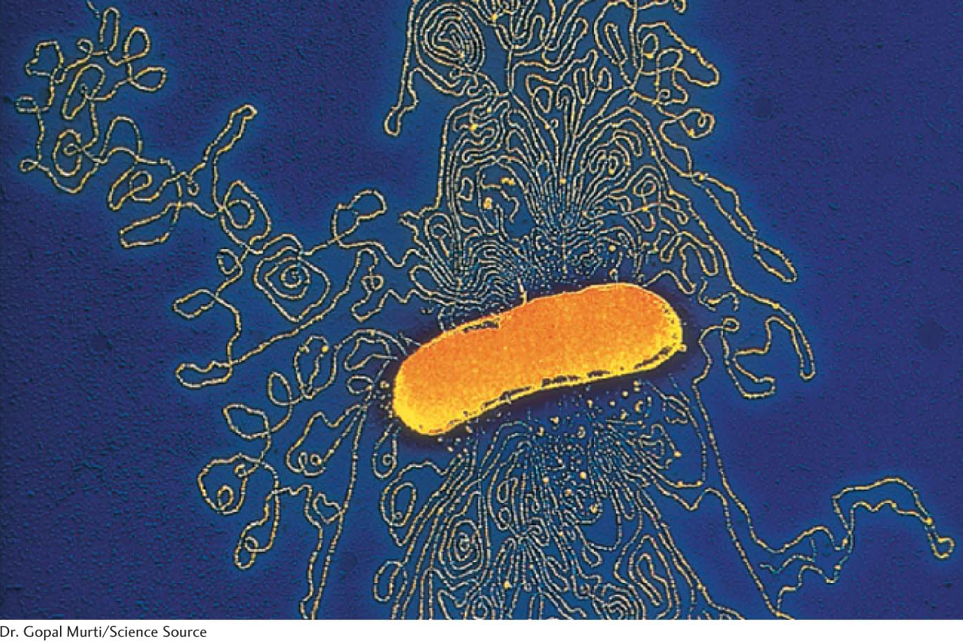
A striking characteristic of naturally occurring DNA molecules is their length. A DNA molecule must comprise many nucleotides to carry the genetic information necessary for even the simplest organisms. For example, the DNA of a virus such as polyoma, which can cause cancer in certain organisms, is 5100 nucleotides in length. The E. coli genome is a single DNA molecule consisting of two strands of 4.6 million nucleotides (Figure 33.9).
DNA molecules from higher organisms can be much larger. The human genome comprises approximately 3 billion base pairs distributed among 24 distinct DNA molecules—
DID YOU KNOW?
Most human cells have 6 billion base pairs of information. All 6 billion base pairs would be 3.6 m in length if all of the molecules were laid end to end. Human beings are composed of approximately 10 trillion cells. If all of this DNA were strung end to end, it would reach to the sun and back about 65 times.
