9.4 An Allosteric Regulator Determines the Oxygen Affinity of Hemoglobin
✓ 8 Identify the key regulators of hemoglobin function.
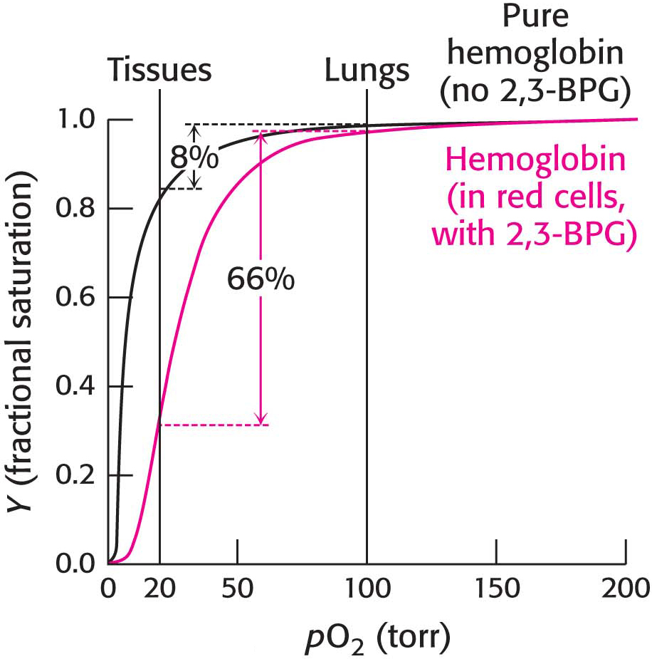
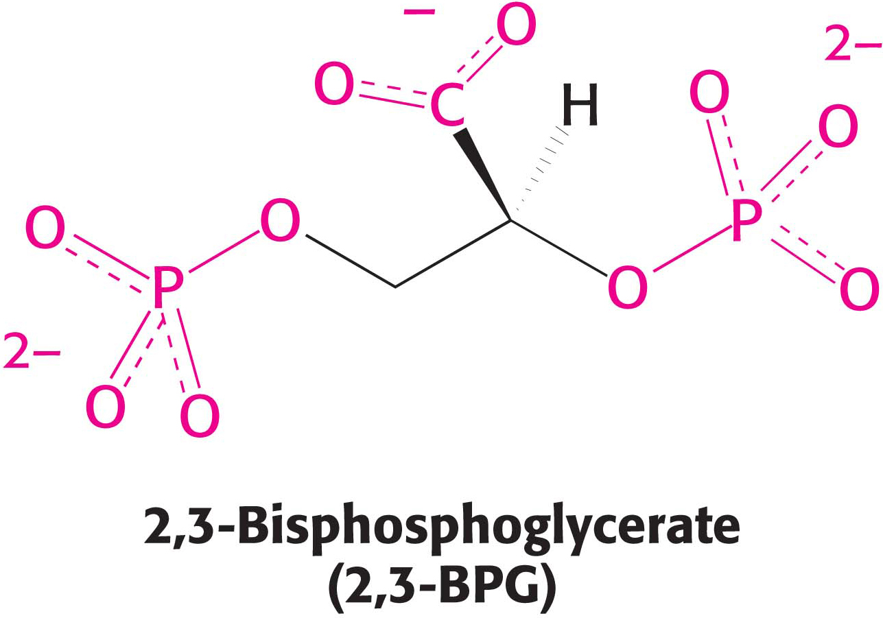
Oxygen binding by hemoglobin is analogous to substrate binding by an allosteric enzyme (Section 7.3). Is the oxygen-binding ability of hemoglobin also affected by regulatory molecules? A comparison of hemoglobin’s behavior in the cell and out is a source of insight into the answer. Hemoglobin can be extracted from red blood cells and fully purified. Interestingly, the oxygen affinity of purified hemoglobin is much greater than that of hemoglobin within red blood cells. The affinity is so great that only 8% of the oxygen would be released to the tissues if hemoglobin in the cell behaved in the way that purified hemoglobin behaves, an amount insufficient to support aerobic tissues. What accounts for the difference between purified hemoglobin and hemoglobin in red blood cells? This dramatic difference is due to the presence, within these cells, of an allosteric regulator molecule—2,3-bisphosphoglycerate (2,3-BPG). This highly anionic compound is present in red blood cells at approximately the same concentration as hemoglobin (~2 mM). 2,3-BPG binds to hemoglobin, reducing its oxygen affinity so that 66% of the oxygen is released instead of a meager 8% (Figure 9.9).
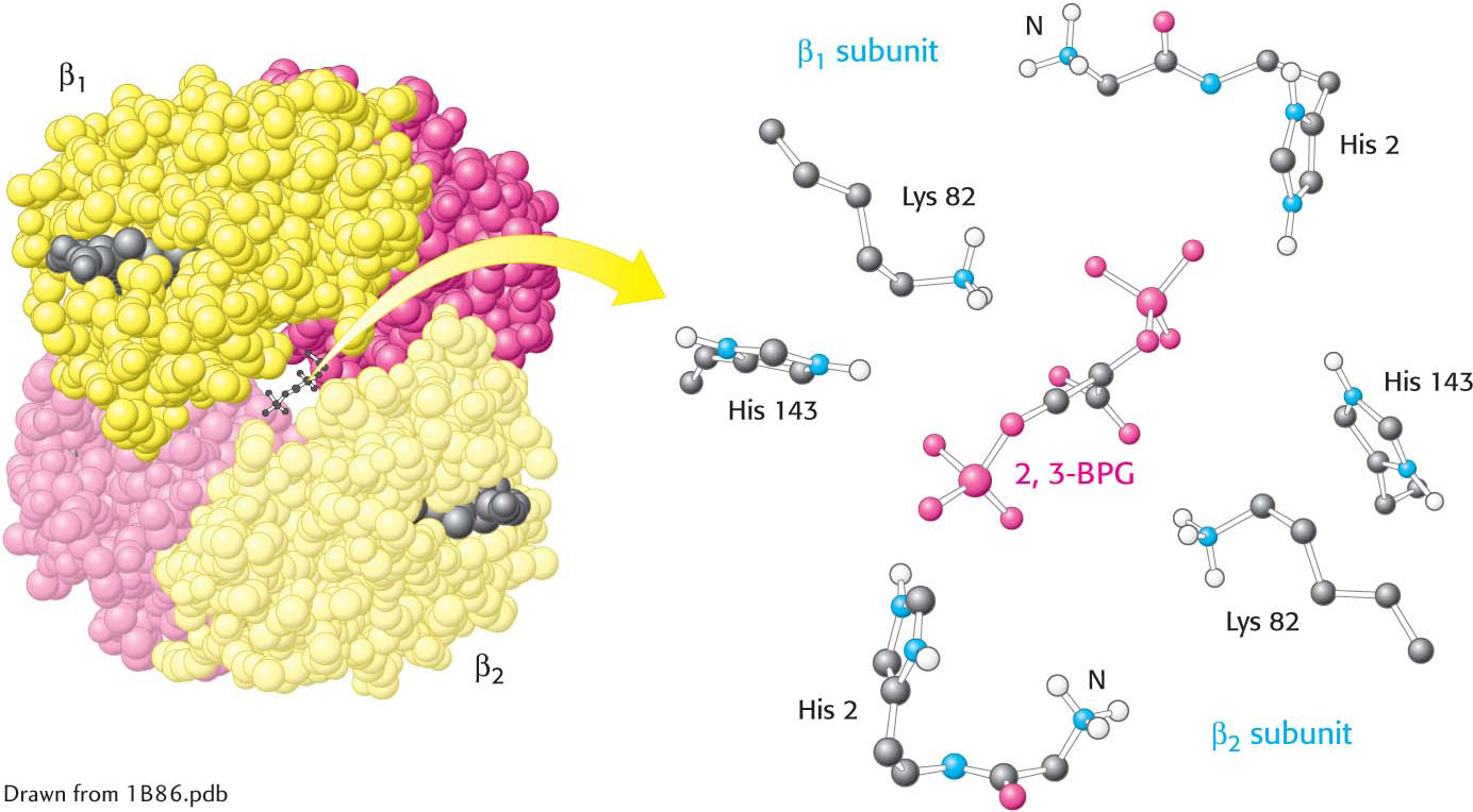
How does 2,3-BPG affect oxygen affinity so significantly? Examination of the crystal structure of deoxyhemoglobin in the presence of 2,3-BPG reveals that a single molecule of 2,3-BPG binds in a pocket, present only in the T form, in the center of the hemoglobin tetramer. The interaction is facilitated by ionic bonds between the negative charges on 2,3-BPG and three positively charged groups of each β chain (Figure 9.10). Thus, 2,3-BPG binds preferentially to deoxyhemoglobin and stabilizes it, effectively reducing the oxygen affinity and leading to the release of oxygen. In order for the structural transition from T to R to take place, the bonds between hemoglobin and 2,3-BPG must be broken and 2,3-BPG must be expelled.
 CLINICAL INSIGHT
Hemoglobin’s Oxygen Affinity Is Adjusted to Meet Environmental Needs
Let us consider another physiological example of the importance of 2,3-BPG binding to hemoglobin. Because a fetus obtains oxygen from the mother’s hemoglobin rather than from the air, fetal hemoglobin must bind oxygen when the mother’s hemoglobin is releasing oxygen. In order for the fetus to obtain enough oxygen to survive in a low-oxygen environment, its hemoglobin must have a high affinity for oxygen. How is the affinity of fetal hemoglobin for oxygen increased?
Page 155
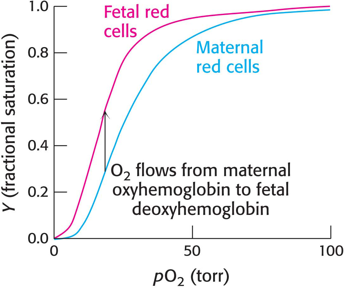
The globin gene expressed by fetuses differs from that expressed by human adults; fetal hemoglobin tetramers include two α chains and two γ chains. The γ chain is 72% identical in amino acid sequence with the β chain. One noteworthy change is the substitution of a serine residue for histidine 143 in the γ chain of the 2,3-BPG-binding site. This change removes two positive charges from the 2,3-BPG-binding site (one from each chain) and reduces the affinity of 2,3-BPG for fetal hemoglobin. Reduced affinity for 2,3-BPG means that the oxygen-binding affinity of fetal hemoglobin is higher than that of the mother’s (adult) hemoglobin (Figure 9.11). This difference in oxygen affinity allows oxygen tobe effectively transferred from the mother’s red blood cells to those of the fetus.
 BIOLOGICAL INSIGHT
Hemoglobin Adaptations Allow Oxygen Transport in Extreme Environments
The bar-headed goose (Figure 9.12) provides another example of hemoglobin adaptations. This remarkable bird migrates over Mt. Everest at altitudes exceeding 9 km (5.6 miles), where the oxygen partial pressure is only 30% of that at sea level. In comparison, consider that human beings climbing Mt. Everest must take weeks to acclimate to the lower pO2 and still usually require oxygen masks. Although the biochemical bases of the bird’s amazing physiological feat are not clearly established, changes in hemoglobin may be important. Compared with its lower-flying cousins, the bar-headed goose has four amino acid changes in its hemoglobin, three in the α chain and one in the β chain. One of the changes in the α chain is proline for alanine, which disrupts a van der Waals contact and facilitates the formation of the R state, thus enabling the protein to more readily bind oxygen.


 CLINICAL INSIGHT
 BIOLOGICAL INSIGHT