Chapter 2. PLANTS
Learning Objectives
General Purpose
Conceptual
- Be able to compare and contrast the major groups within the kingdom Plantae.
- Be able to describe the major evolutionary trends in the kingdom Plantae.
- Be able to differentiate between the sporophyte and gametophyte generations.
Procedural
- Be able to compare and contrast the major groups within the kingdom Plantae.
- Be able to construct a phylogenetic tree of the kingdom Plantae.
Background Information
Plants are eukaryotic, multicellular, autotrophic organisms that use photosynthesis to convert light energy to chemical energy. Most forms are terrestrial. Land plants generally have complex, multicellular plant bodies that are specialized for a variety of functions. Specialized structures have evolved for protection of the vulnerable stages of sexual reproduction. The plant body is often covered with a waxy cuticle that prevents desiccation.
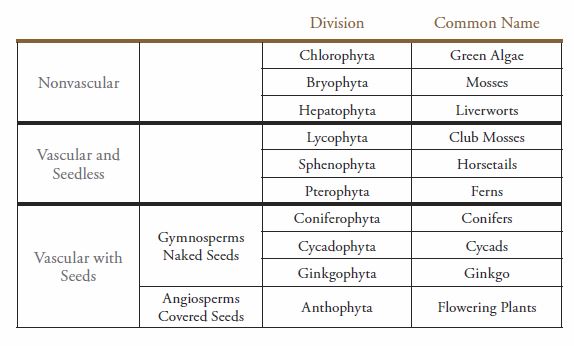
While plants present a large array of diversity, there are a couple of themes that run throughout this kingdom (Table 10-2). These themes include changes in size, changes in dependence on water, changes in vascular tissue, and changes in the dominance of the stages of the life cycle.
Table 10-2. Classification of land plants.
Plant Life Cycle
Land plants have a common sexual reproductive life cycle called alternation of generations, in which plants alternate between a haploid gametophyte generation and a diploid sporophyte generation (Figure 10-13).
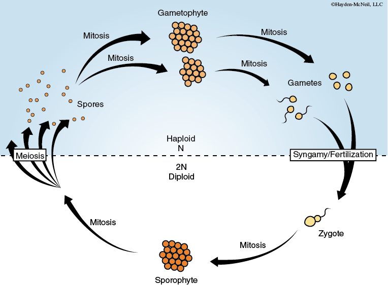
These two generations differ in their morphology. In all land plants except the bryophytes (mosses) and hepatophytes (liverworts), the diploid sporophyte generation is the dominant (more conspicuous) generation. The sporophyte of the vascular plants can be tall and have large leaves, although in many vascular plants the sporophyte is low growing. The sporophyte generation undergoes meiosis to produce haploid spores. The spores germinate to produce the haploid gametophyte, which is independent of the sporophyte in nonvascular and seedless vascular plants, but dependent on the sporophyte in the seed plants. The gametophyte produces haploid gametes by mitosis. Fertilization (fusion of the gametes) results in a diploid zygote, which is the first stage of the diploid sporophyte generation.
Note that both gametes and spores are haploid in this life cycle. The difference between these two cells is that gametes fuse with other gametes to form the zygote and restore the diploid number, while spores germinate to form a new haploid gametophyte plant.
Exercise 1. Create a Phylogenetic Tree for the Plant Kingdom
PROCEDURE
- Use the in-lab cards to create a tree for the plant kingdom. Sketch the tree in your laboratory notebook. Use this tree during the rest of the class and add characteristics.
Exercise 2. Division Chlorophyta
Materials
Microscope
Depression slide
Fresh, preserved or prepared specimens of various members of Division Chlorophyta
PROCEDURE
- Use a compound microscope to observe living materials or prepared slides of the filamentous alga Spirogyra sp.
- Record any observations in your laboratory notebook.
- In your laboratory notebook make a sketch of one or more views of the specimens you are observing.
- Observe the preserved specimen of Ulva sp., commonly called sea lettuce. This multicellular alga is often found on rocks or docks in marine and brackish water.
- In your laboratory notebook, make a sketch of one or more views of the specimens you are observing.
- Prepare a wet mount of Chlamydomonas sp. Use the compound microscope to observe the single-celled motile Chlamydomonas.
- Record any observations in your laboratory notebook including swimming behavior.
- In your laboratory notebook make a sketch of one or more views of the specimens you are observing.
Exercise 3. Division Bryophyta
Materials
Fresh specimens of members of Division Bryophyta
PROCEDURE
- Examine living colonies of mosses at the demonstration station. Usually you will find the two generations, gametophyte and sporophyte, growing together.
- Identify the leafy gametophytes and the dependent sporophytes, which appear as elongated structures growing above them.
- Record any observations in your laboratory notebook.
- In your laboratory notebook, make a sketch of one or more views of the specimens you are observing.
- Frequently, the spores for organisms in this division are wind-dispersed. If you have not already done so, view video of spores being forcibly expelled.
Exercise 4. Division Hepatophyta
Materials
Microscope
Fresh and prepared specimens of members of Division Hepatophyta
PROCEDURE
- Examine examples of liverworts at the demonstration station. Liverworts have a flat thallus (plant body).
- Record any observations in your laboratory notebook.
- In your laboratory notebook, make a sketch of one or more views of the specimens you are observing. Note the rhizoids, root-like extensions on the lower surface. On the upper surface of the thallus you should see circular cups called gemmae cups, which contain flat disks of green tissue called gemmae. The gemmae are washed out of the cups when it rains, and they grow into new, genetically identical liverworts.
- Use the microscope to examine a cross-section slide of liverworts thallus.
- Record any observations in your laboratory notebook.
- In your laboratory notebook, make a sketch of one or more views of the specimens you are observing.
Exercise 5. Division Lycophyta (Seedless Vascular Plants)
Materials
Microscope
Fresh, preserved and prepared specimens of members of Division Lycophyta
PROCEDURE
- Examine living club mosses, Selaginella and Lycopodium.
- Record any observations in your laboratory notebook. Make note of the type of branching pattern in the specimens. If they are dichotomously branched, then the branches would split in two, appearing to form a Y.
- In your laboratory notebook, make a sketch of one or more views of the specimens you are observing.
- Examine the preserved strobili of Selaginella.
- Record any observations in your laboratory notebook. Note the round sporangia clustered in sporophylls (leaf-like structures) at the tip of the stem.
- In your laboratory notebook, make a sketch of one or more views of the specimens you are observing.
- Use the microscope to observe the prepared slide of a long section through the strobilus of Selaginella. Begin your observations at low power. In your laboratory notebook make a sketch of one or more views of the specimens you are observing.
Exercise 6. Division Sphenophyta
Materials
Fresh specimens of members of Division Sphenophyta
PROCEDURE
- Examine examples of horsetails at the demonstration station.
- Record any observations in your laboratory notebook. Note the ribs and ridges in the stem. Also examine the nodes, or joints, along the stem where branches and leaves may occur in some species. Locate the strobili. These are clusters of sporangia that produce spores.
- In your laboratory notebook, make a sketch of the overall structure of the horsetails and label structures where appropriate.
Exercise 7. Division Pterophyta
Materials
Dissecting microscope
Depression slide
Fresh specimens of members of Division Pterophyta
PROCEDURE
- Examine a living fern sporophyte.
- Record any observations in your laboratory notebook. Note the deeply dissected leaves, which arise from an underground stem called a rhizome, which functions like a root to anchor the plant. Observe the dark spots or sori (singular sorus), which are clusters of sporangia, on the underside of some leaves.
- In your laboratory notebook, make a sketch of the overall structure of the fern sporophyte and label structures where appropriate.
- Make a wet mount of sporangia and examine the sporangia using a dissecting microscope.
- Record any observations in your laboratory notebook. You should find the stalked sporangia in various stages of development.
- In your laboratory notebook, make a sketch of the specimens.
Exercise 8. Gymnosperms (Seed Plants with Naked Seeds)
Materials
Fresh specimens of members of Gymnosperms
PROCEDURE
- Examine examples of all divisions of gymnosperms at the demonstration station.
- Record any observations in your laboratory notebook and be able to recognize their representatives. Note any significant ecological and economic role for these plants.
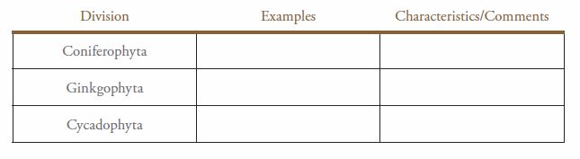
- In you laboratory notebook, make a table like the one shown below (Table 10-3) to record your observations of the different divisions of Gymnosperms.
Table 10-3. Divisions of Gymnosperms.
Exercise 9. Anthophyta (Seed Plants with Covered Seeds)
Materials
Fresh specimens of members of Anthophyta
PROCEDURE
- Examine examples of all divisions of Anthophyta at the demonstration station.
- Record any observations in your laboratory notebook and be able to recognize their representatives. Note any significant ecological and economic role for these plants.
- In your laboratory notebook, make a list of the features that separate Anthophyta from the Gymnosperms.
Exercise 10. Summary of Plant Characteristics
PROCEDURE
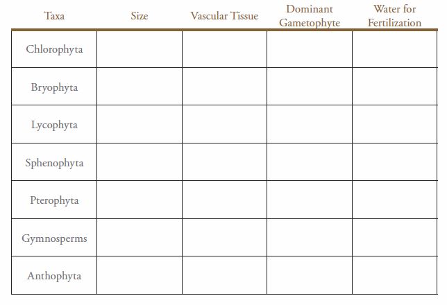
- In your laboratory notebook, make a table similar to the one shown below (Table 10-4).
Complete the table using the observations of the different plant representatives.
Table 10-4. Summary of plant characteristics.
Post-Lab Quiz
Proceed to the Post-Lab Quiz