Chapter 1. Tour of the Cell
Tour of the Cell
Tour of the Cell
by Anthony Kain and Grishma Acharya
CELLS
The Fundamental Units of Life
All living organisms are composed of matter that is structured with increasing complexity:
atoms → molecules → macromolecules → organelles → cells → tissues → organs → systems → organism
The cell is the first of these units to contain everything required for life: the ability to grow (metabolize) and reproduce and contain heritable material. As such, it is the fundamental unit of life and most organisms consist of a single cell (most organisms are single-celled prokaryotes). While living organisms from all taxonomic kingdoms differ vastly in anatomy and morphology, the cells that make up living organisms are surprisingly similar, especially at the molecular level.
The Prokaryotic Cell
The Prokaryotic Cell
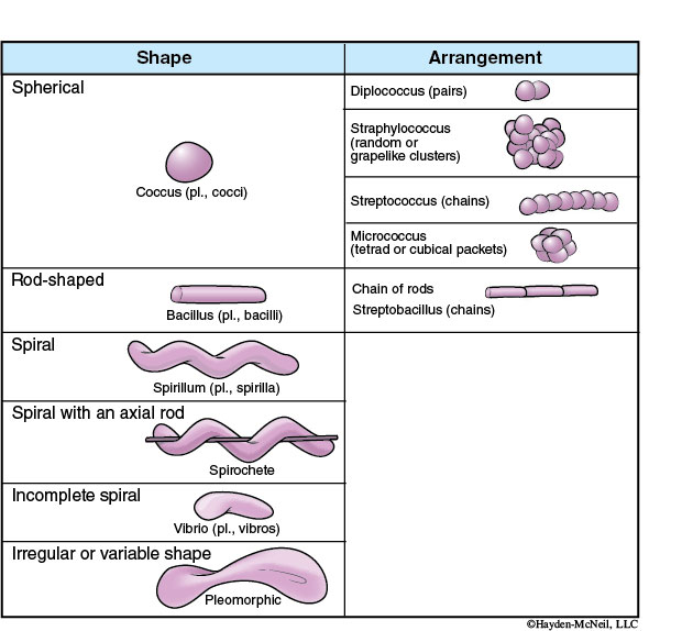
Prokaryotes (Bacteria and Archaea) show incredible diversity in their metabolism (the chemical reactions they perform) but have a simple morphology and are extremely small. Like all cells, prokaryotes have a plasma membrane separating them from the environment. Prokaryotes have a nucleoid region where the cell’s DNA is packaged in a circular chromosome. Prokaryotes also have cell walls that function in a similar way to those in plants and fungi; however, prokaryotic cell walls are usually made of a compound called peptidoglycan instead of cellulose. Some prokaryotes have an additional capsule outside their cell wall. Others have flagella which, like in sperm, move the cell. However, at the molecular level, prokaryotic flagella are totally different from those in sperm. Instead of a whipping motion, prokaryotes spin their flagella around like a propeller.
Bacteria come in three basic shapes: spherical, rod-shaped, and spiral (Table 4-1).
The Eukaryotic Cell
The Eukaryotic Cell
The defining feature of eukaryotes is the presence of a nucleus enclosed within a double-membrane nuclear envelope. A darkened region called the nucleolus is the part of the nucleus where ribosome synthesis occurs. Eukaryotes show much more diversity in their cell structure than do prokaryotes; multicellular eukaryotes, like plants and ourselves, have lots of organelles (discrete cell structures), but all eukaryotes have a nucleus, a plasma membrane, ribosomes, and a cytoskeleton. Eukaryotic cells tend to be larger than prokaryotic cells, and the cytoskeleton provides necessary structural support for these larger cells as well as helping with cell motility and the transport of materials throughout the cell. Specific structures involved in cell motility are cilia, flagella, and pseudopodia.
Cillium
Cilium (plural: cilia) is a slender, microscopic, hair-like structure or organelle that extends from the surface of some cells and certain organisms (multiple or single). Cilia can be divided into two subcategories in different cells—motile cilia (for locomotion) or primary cilia (non-motile, used for sensory purposes), depending on what the cell/organism uses the cilia for. Today we will notice how cilia are used by paramecia for movement or to escape from predators, as well as for sweeping food into their bodies.
Flagellum
Flagella are appendages that protrude from the cell body of certain prokaryotic and eukaryotic cells. They are structurally very similar to cilia, but larger. We will notice flagella on an organism named Euglena today. This flagellated protozoan uses a flagellum to move.
Pseudopodia
Pseudopodia (meaning “false feet”) are another means of locomotion for cells. They are extensions of the cytoplasm toward which the rest of the cytoplasm tends to flow. The Amoeba is the most notable organism that uses pseudopodia as a means of motion as well food consumption.
Many eukaryotic organelles are membrane-bound compartments that allow the cell to compartmentalize various chemical reactions. This is a huge advantage because each organelle can specialize in certain types of chemical reactions and optimize conditions for those reactions by providing a microenvironment with the proper morphology, pH, substrate concentrations, etc. For example, both mitochondria and chloroplasts have a lot of internal membranes, which provide space for electron transport chain proteins and separate compartments with different pHs. Once again we see the correlation between structure and function.
Cells can be thought of as miniature factories in which there are several departments (organelles). Each department is responsible for a specific duty that ultimately contributes to a specific objective. Among these functions are: obtaining (heterotrophs like animals and fungi) or generating (autotrophs like plants) food, metabolism and energy production, waste management, and production or modification of macromolecules like proteins/enzymes.
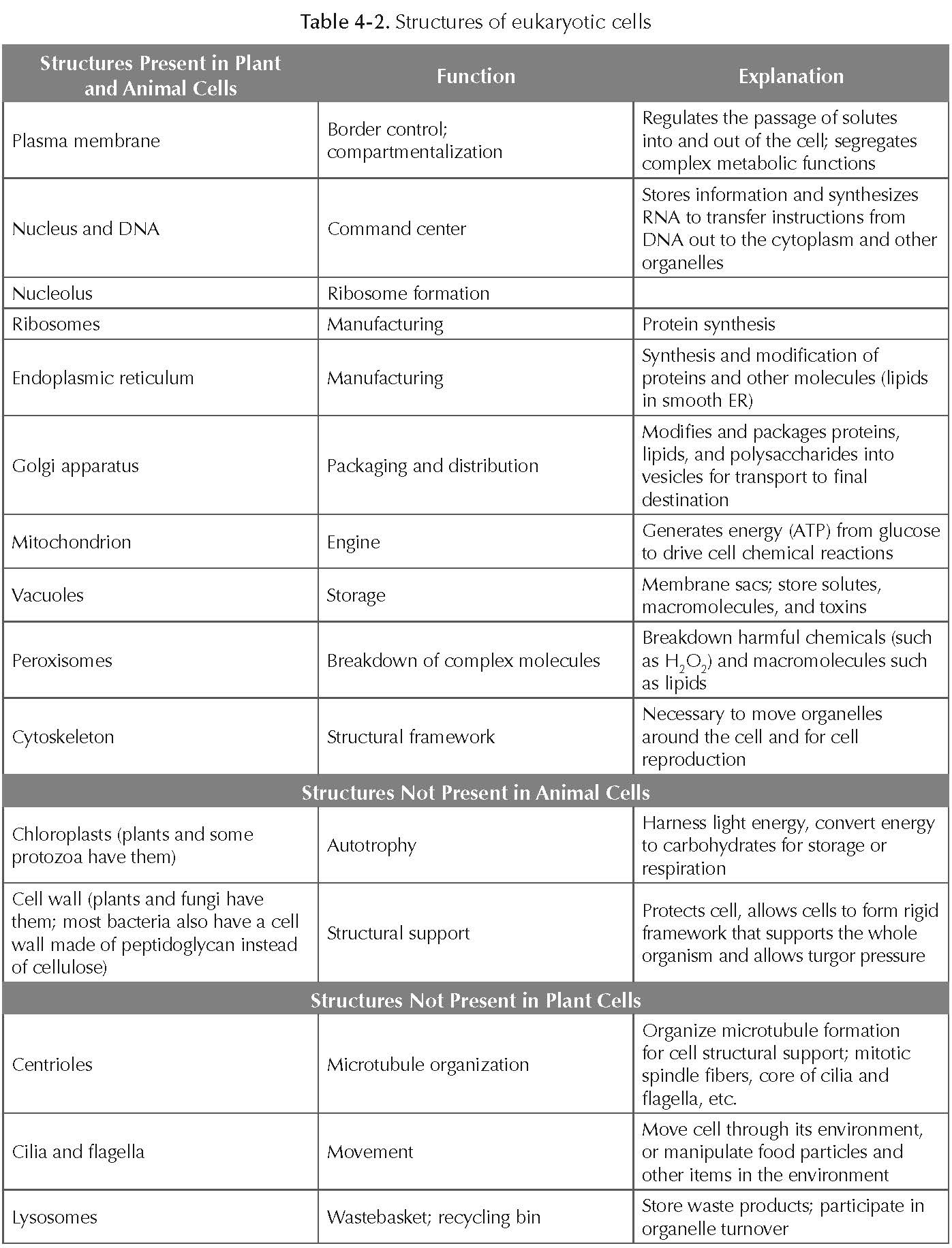
The following table contains a list of cellular structures followed by a description of their function and a brief explanation. You should be able to identify these structures on cell diagrams and give their functions. You may want to refer to the cell models available in lab or to the internet resources listed at the end of this lab for further information.
During the lab you will observe unicellular (one-celled) eukaryotic living organisms named Protozoa. Live specimens will include Euglena, Paramecium, and Amoeba. We will be focusing on three different types of cell motility these organisms use.
Euglena
Euglena
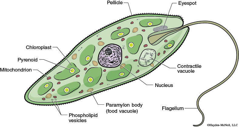
This is a single-celled, freshwater protozoan. This unique organism contains chloroplasts with chlorophyll and can photosynthesize as a plant. It also has a flagellum to move, and this is usually a characteristic of an animal cell.
Paramecium
Paramecium
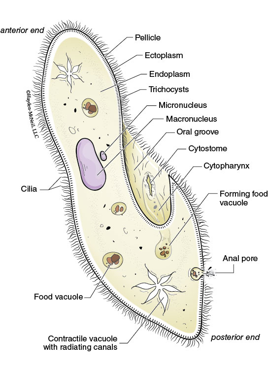
The Paramecium is an oval, slipper-shaped organism, rounded at the front/top and pointed at the back/bottom. They have many tiny hairs called cilia that assist in the mobility functions of this organism. Paramecia are famous for their predator-prey relationship with heterotrophic organisms called Didinium that prey upon paramecia. When a Didinium finds a Paramecium, it ejects poison darts (trichocysts) and then proceeds to engulf its prey. Although paramecia are larger than they are, Didinium are voracious eaters and will be ready to hunt for another meal after only a few hours. Upon encounter with a negative stimulus (such as the predator Didinium), Paramecium is capable of rotating 360 degrees to find an escape route.
Amoeba
Amoeba
Another organism from the protozoan group, Amoeba is a unicellular organism always changing its shape that is famous for its movement techniques. Amoeba is mobile by virtue of tentacle-like protrusions commonly called “false feet” (scientifically: pseudopodia). These pseudopodia are used for mobility as well as to phagocytize smaller unicellular organisms by enveloping them inside the cell’s cytoplasm in a food vacuole; thereafter, they are slowly broken down by enzymes.
Microscope
Microscope
A microscope is the main tool to observe and study cells. There are several different kinds of microscopes that are used to study cells. Light microscopes (found in this lab) focus visible light through refractory lenses to magnify an image. Figure 4-2 shows a typical compound light microscope; this microscope gets its name from the fact that it uses multiple lenses (ocular and objective) and light to magnify specimens. Electron microscopes have approximately 1,000 times the magnifying power of light microscopes and are used to study the ultrastructure of cells. These microscopes use a beam of electrons (instead of light) to penetrate a thin section of a specimen and can reveal extremely small organelles that cannot be viewed using a light microscope.
Proper Handling and Use of the Compound Light Microscope
Please handle these microscopes with care. The proper way to carry a light microscope is to hold it with one hand around the arm of the microscope while supporting the microscope with your other hand under the base. Plug the microscope into an electrical outlet and turn on the light source. The amount of light passing through your specimen can be controlled in two ways. The intensity of the light source can be adjusted by the rheostat located near the base of the microscope. The iris diaphragm and condenser can be used to control the amount of light passing through your specimen; use these to optimize your view of your specimen. Do not slide the microscope on the lab bench; this may damage the microscope.