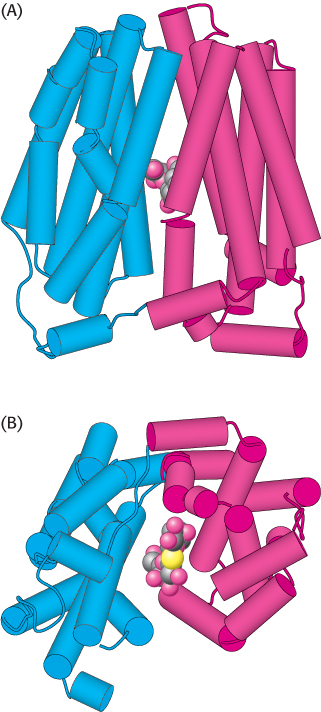
Structure of lactose permease with a bound lactose analog. The amino-terminal half of the protein is shown in blue and the carboxyl-terminal half in red. (A) Side view. (B) Bottom view (from inside the cell). Notice that the structure consists of two halves that surround the sugar and are linked to one another by only a single stretch of polypeptide. [Drawn from 1PV7.pdb.]

