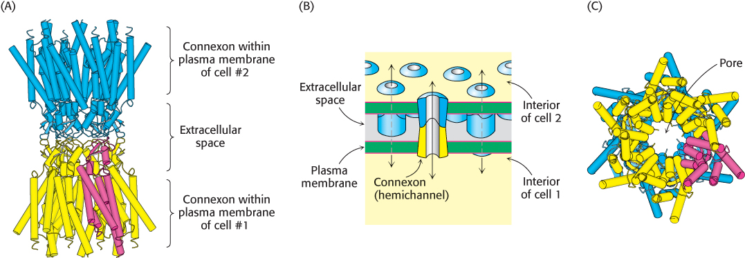
Structure of a gap junction. (A) Six connexins join to form a connexon, or hemichannel, within the plasma membrane (yellow). A single connexin monomer is highlighted in red. The extracellular region of one connexon binds to the same region of a connexon from another cell (blue), forming a complete gap junction. (B) Schematic view of the gap junction, oriented in the same direction as in (A). (C) A bottom-