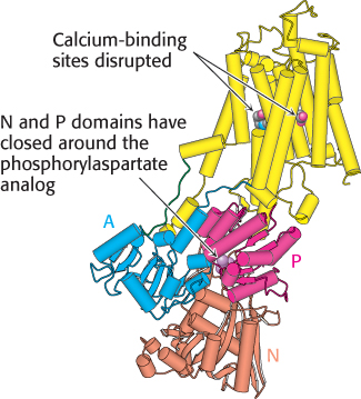
Conformational changes associated with calcium pumping. This structure was determined in the absence of bound calcium but with a phosphorylaspartate analog present in the P domain. Notice how different this structure is from the calcium-bound form shown in Figure 13.3: both the transmembrane part (yellow) and the A, P, and N domains have substantially rearranged. [Drawn from 1WPG.pdb.]