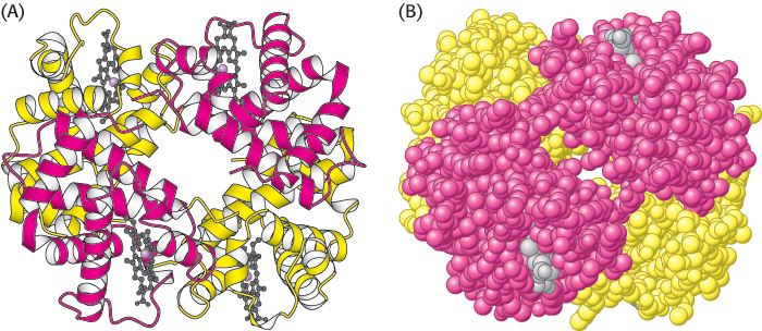
The α2β2 tetramer of human hemoglobin. The structure of the two α subunits (red) is similar to but not identical with that of the two β subunits (yellow). The molecule contains four heme groups (gray with the iron atom shown in purple). (A) The ribbon diagram highlights the similarity of the subunits and shows that they are composed mainly of α helices. (B) The space-
[Drawn from 1A N.pdb.]