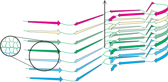
A model of the human prion protein amyloid. A detailed model of a human prion amyloid fibril deduced from spin labeling and electron paramagnetic resonance (EPR) spectroscopy studies shows that protein aggregation is due to the formation of large parallel β sheets. The black arrow indicates the long axis of the fibril.
[N. J. Cobb, F. D. Sönnichsen, H. Mchaourab, and W. K. Surewicz. Proc. Natl. Acad. Sci. U.S.A. 104: 18946–