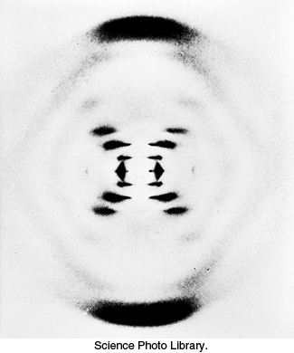
X-ray diffraction photograph of a hydrated DNA fiber. When crystals of a biomolecule are irradiated with x-rays, the x-rays are diffracted and these diffracted x-rays are seen as a series of spots, called reflections, on a screen behind the crystal. The structure of the molecule can be determined by the pattern of the reflections (Section 3.5). In regard to DNA crystals, the central cross is diagnostic of a helical structure. The strong arcs on the meridian arise from the stack of nucleotide bases, which are 3.4 Å apart.
[Science Photo Library.]