Watson–Crick model of double-helical DNA. (A) Side view. Adjacent bases are separated by 3.4 Å. The structure repeats along the helical axis (vertical) at intervals of 34 Å, which corresponds to approximately 10 nucleotides on each chain. (B) Axial view, looking down the helix axis, reveals a rotation of 36° per base and shows that the bases are stacked on top of one another
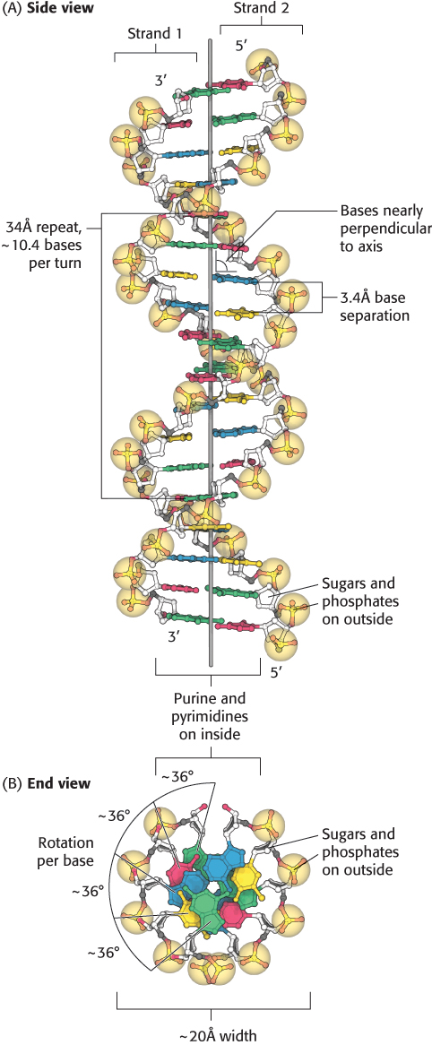
[Source: J. L. Tymoczko, J. Berg, and L. Stryer, Biochemistry: A Short Course, 2nd ed. (W. H. Freeman and Company, 2013), Fig. 33.11.].