22.2 The Use of Fatty Acids as Fuel Requires Three Stages of Processing
Tissues throughout the body gain access to the lipid energy reserves stored in adipose tissue through three stages of processing. First, the lipids must be mobilized. In this process, triacylglycerols are degraded to fatty acids and glycerol, which are released from the adipose tissue and transported to the energy-
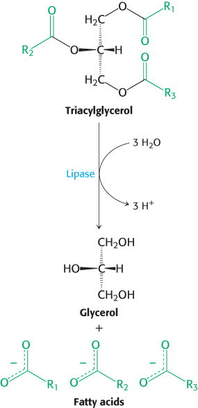
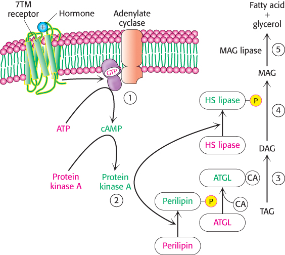
Triacylglycerols are hydrolyzed by hormone-stimulated lipases
Consider someone who has just awakened from a night’s sleep and begins a bout of exercise. Glycogen stores will be low, but lipids are readily available. How are these lipid stores mobilized?
Before fats can be used as fuels, the triacylglycerol storage form must be hydrolyzed to yield isolated fatty acids. This reaction is catalyzed by a hormonally controlled lipase. Under the physiological conditions facing an early-
648
 If the coactivator required by ATGL is missing or defective, a rare condition (incidence unknown) called Chanarin-
Free fatty acids and glycerol are released into the blood
Fatty acids are not soluble in aqueous solutions. In order to reach tissues that require fatty acids, t he released fatty acids bind to the blood protein albumin, which delivers them to tissues in need of fuel.
Glycerol formed by lipolysis is absorbed by the liver and phosphorylated. It is then oxidized to dihydroxyacetone phosphate, which is isomerized to glyceraldehyde 3-
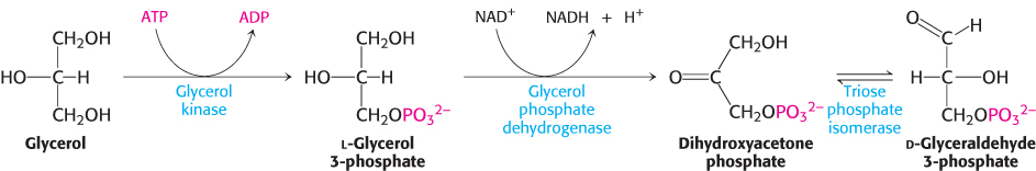
Hence, glycerol can be converted into pyruvate or glucose in the liver, which contains the appropriate enzymes (Figure 22.7). The reverse process can take place by the reduction of dihydroxyacetone phosphate to glycerol 3-

Fatty acids are linked to coenzyme A before they are oxidized
Fatty acids separate from the albumin in the blood stream and diffuse across the cell membrane with the assistance of transport proteins. In the cell, fatty acids are shuttled about in association with fatty-
649
Fatty acid oxidation occurs in the mitochondrial matrix, but in order to enter the mitochondria, the fatty acids are first activated through the formation of a thioester linkage to coenzyme A. Adenosine triphosphate (ATP) drives the formation of the thioester linkage between the carboxyl group of a fatty acid and the sulfhydryl group of coenzyme A. This activation reaction takes place on the outer mitochondrial membrane, where it is catalyzed by acyl CoA synthetase (also called fatty acid thiokinase).
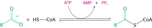
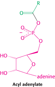
Acyl CoA synthetase accomplishes the activation of a fatty acid in two steps. First, the fatty acid reacts with ATP to form an acyl adenylate. In this mixed anhydride, the carboxyl group of a fatty acid is bonded to the phosphoryl group of AMP. The other two phosphoryl groups of the ATP substrate are released as pyrophosphate. In the second step, the sulfhydryl group of coenzyme A attacks the acyl adenylate, which is tightly bound to the enzyme, to form acyl CoA and AMP.

These partial reactions are freely reversible. In fact, the equilibrium constant for the sum of these reactions is close to 1. One high-

This reaction is quite favorable because the equivalent of two molecules of ATP is hydrolyzed, whereas only one high-
 Another motif recurs in this activation reaction. The enzyme-
Carnitine carries long-chain activated fatty acids into the mitochondrial matrix
Fatty acids are activated on the outer mitochondrial membrane, whereas they are oxidized in the mitochondrial matrix. A special transport mechanism is needed to carry activated long-
650
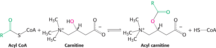
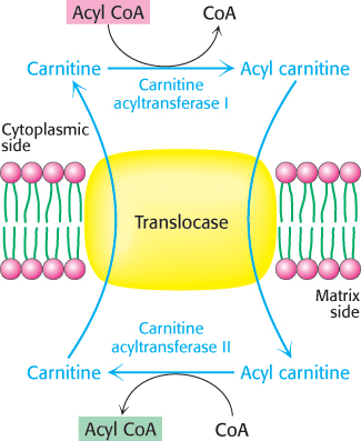
Acyl carnitine is then shuttled across the inner mitochondrial membrane by a translocase (Figure 22.8). The acyl group is transferred back to coenzyme A on the matrix side of the membrane. This reaction, which is catalyzed by carnitine acyltransferase II (carnitine palmitoyl transferase II), is simply the reverse of the reaction that takes place in the cytoplasm. The reaction is thermodynamically feasible because of the zwitterionic nature of carnitine. The O-acyl link in carnitine has a high group-
A number of diseases have been traced to a deficiency of carnitine, the transferase, or the translocase. Inability to synthesize carnitine may be a contributing factor to the development of autism in males. The symptoms of carnitine deficiency range from mild muscle cramping to severe weakness and even death. In general, muscle, kidney, and heart are the tissues primarily impaired. Muscle weakness during prolonged exercise is a symptom of a deficiency of carnitine acyltransferases because muscle relies on fatty acids as a long-
Acetyl CoA, NADH, and FADH2 are generated in each round of fatty acid oxidation

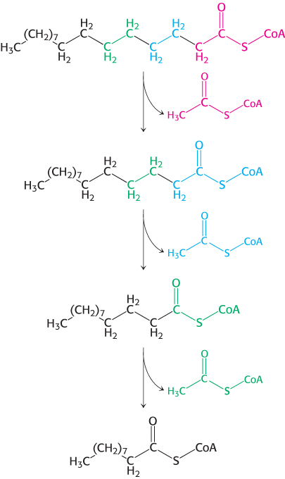
A saturated acyl CoA is degraded by a recurring sequence of four reactions: oxidation by flavin adenine dinucleotide (FAD), hydration, oxidation by NAD+, and thiolysis by coenzyme A (Figure 22.9). The fatty acid chain is shortened by two carbon atoms as a result of these reactions and FADH2, NADH, and acetyl CoA are generated. Because oxidation takes place at the β carbon atom, this series of reactions is called the β-oxidation pathway.
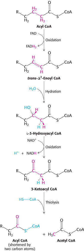
The first reaction in each round of degradation is the oxidation of acyl CoA by an acyl CoA dehydrogenase to give an enoyl CoA with a trans double bond between C-

As in the dehydrogenation of succinate in the citric acid cycle, FAD rather than NAD+ is the electron acceptor because the ΔG for this reaction is insufficient to drive the reduction of NAD+. Electrons from the FADH2 prosthetic group of the reduced acyl CoA dehydrogenase are transferred to a second flavoprotein called electron-
651

The next step is the hydration of the double bond between C-

The hydration of enoyl CoA is stereospecific. Only the l isomer of 3-
The hydration of enoyl CoA is a prelude to the second oxidation reaction, which converts the hydroxyl group at C-

The preceding reactions have oxidized the methylene group at C-

Table 22.1 summarizes the reactions in fatty acid oxidation.
|
Step |
Reaction |
Enzyme |
|---|---|---|
|
1 |
|
Acyl CoA synthetase (also called fatty acid thiokinase and fatty acid:CoA ligase)* |
|
2 |
|
Carnitine acyltransferase (also called carnitine palmitoyl transferase) |
|
3 |
|
Acyl CoA dehydrogenases (several isozymes having different chain- |
|
4 |
|
Enoyl CoA hydratase (also called crotonase or 3- |
|
5 |
|
l-3- |
|
6 |
|
β-Ketothiolase (also called thiolase) |
|
*An AMP- |
||


The shortened acyl CoA then undergoes another cycle of oxidation, starting with the reaction catalyzed by acyl CoA dehydrogenase (Figure 22.10). Fatty acid chains containing from 12 to 18 carbon atoms are oxidized by the long-
652
The complete oxidation of palmitate yields 106 molecules of ATP
We can now calculate the energy yield derived from the oxidation of a fatty acid. In each reaction cycle, an acyl CoA is shortened by two carbon atoms, and one molecule each of FADH2, NADH, and acetyl CoA are formed.

The degradation of palmitoyl CoA (C16-acyl CoA) requires seven reaction cycles. In the seventh cycle, the C4-ketoacyl CoA is thiolyzed to two molecules of acetyl CoA. Hence, the stoichiometry of the oxidation of palmitoyl CoA is

Approximately 2.5 molecules of ATP are generated when the respiratory chain oxidizes each of these NADH molecules, whereas 1.5 molecules of ATP are formed for each FADH2 because their electrons enter the chain at the level of ubiquinol. Recall that the oxidation of acetyl CoA by the citric acid cycle yields 10 molecules of ATP. Hence, the number of ATP molecules formed in the oxidation of palmitoyl CoA is 10.5 from the seven FADH2, 17.5 from the seven NADH, and 80 from the eight acetyl CoA molecules, which gives a total of 108. The equivalent of 2 molecules of ATP is consumed in the activation of palmitate, in which ATP is split into AMP and 2 molecules of orthophosphate. Thus, the complete oxidation of a molecule of palmitate yields 106 molecules of ATP.