33.2 Taste Is a Combination of Senses That Function by Different Mechanisms

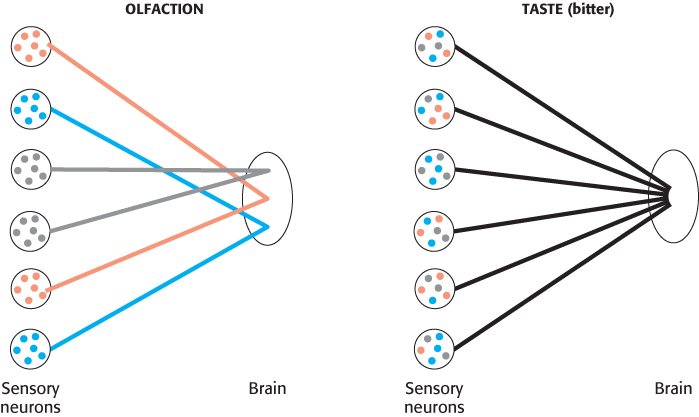
The inability to taste food is a common complaint when nasal congestion reduces the sense of smell. Thus, smell greatly augments our sense of taste (also known as gustation), and taste is, in many ways, the sister sense to olfaction. Nevertheless, the two senses differ from each other in several important ways. First, we are able to sense several classes of compounds by taste that we are unable to detect by smell; salt and sugar have very little odor, yet they are primary stimuli of the gustatory system. Second, whereas we are able to discriminate thousands of odorants, discrimination by taste is much more modest. Five primary tastes are perceived: bitter, sweet, sour, salty, and umami (the taste of glutamate and aspartate from the Japanese word for “deliciousness”). These five tastes serve to classify compounds into potentially nutritive and beneficial (sweet, salty, umami) or potentially harmful or toxic (bitter, sour). Tastants (the molecules sensed by taste) are quite distinct for the different groups (Figure 33.10).

Figure 33.10: Examples of tastant molecules. Tastants fall into five groups: sweet, salty, umami, bitter, and sour.
Figure 33.11: A taste bud. Each taste bud contains sensory neurons that extend microvilli to the surface of the tongue, where they interact with tastants.

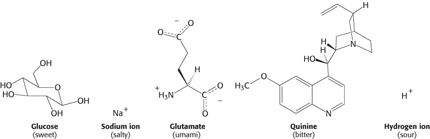
The simplest tastant, the hydrogen ion, is perceived as sour. Other simple ions, particularly sodium ion, are perceived as salty. The taste called umami is evoked by the amino acids glutamate and aspartate, the former often encountered as the flavor enhancer monosodium glutamate (MSG). In contrast, tastants perceived as sweet and, particularly, bitter are extremely diverse. Many bitter compounds are alkaloids or other plant products, many of which are toxic. However, they do not have any common structural elements or other common properties. Carbohydrates such as glucose and sucrose are perceived as sweet, as are other compounds including some simple peptide derivatives, such as aspartame, and even some proteins.

967

These differences in specificity among the five tastes are due to differences in their underlying biochemical mechanisms. The sense of taste is, in fact, a number of independent senses all utilizing the same organ, the tongue, for their expression.

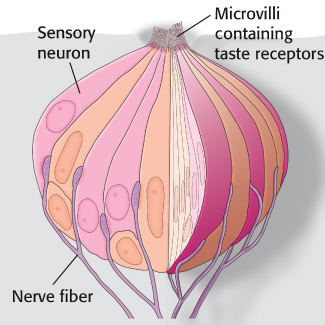
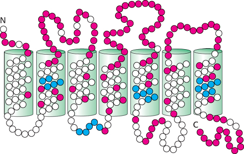
Tastants are detected by specialized structures called taste buds, which contain approximately 150 cells, including sensory neurons (Figure 33.11). Fingerlike projections called microvilli, which are rich in taste receptors, project from one end of each sensory neuron to the surface of the tongue. Nerve fibers at the opposite end of each neuron carry electrical impulses to the brain in response to stimulation by tastants. Structures called taste papillae contain numerous taste buds.

Sequencing of the human genome led to the discovery of a large family of 7TM bitter receptors

Figure 33.12: Expression of gustducin in the tongue. (A) A section of tongue stained with a fluorescent antibody reveals the position of the taste buds. (B) The same region stained with an antibody directed against gustducin reveals that this G protein is expressed in taste buds.

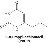
Just as in olfaction, a number of clues pointed to the involvement of G proteins and, hence, 7TM receptors in the detection of bitter and sweet tastes. The evidence included the isolation of a specific G-protein α subunit termed gustducin, which is expressed primarily in taste buds (Figure 33.12). How could the 7TM receptors be identified? The ability to detect some compounds depends on specific genetic loci in both human beings and mice. For instance, the ability to taste the bitter compound 6-n-propyl-2-thiouracil (PROP) was mapped to a region on human chromosome 5 by comparing DNA markers of persons who vary in sensitivity to this compound.

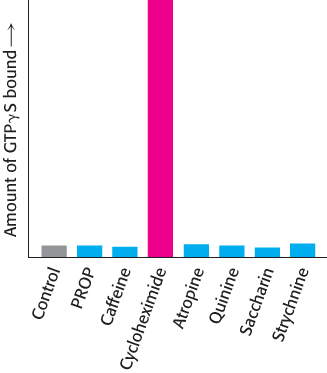
This observation suggested that this region might encode a 7TM receptor that responded to PROP. Approximately 450 kilobases in this region had been sequenced early in the human genome project. This sequence was searched by computer for potential 7TM-receptor genes, and, indeed, one was detected and named T2R1. Additional database searches detected approximately 30 sequences similar to T2R1 in the human genome. The encoded proteins are between 30 and 70% identical with T2R1 (Figure 33.13).

Figure 33.13: Conserved and variant regions in bitter receptors. The bitter receptors are members of the 7TM-receptor family. Strongly conserved residues characteristic of this protein family are shown in blue, and highly variable residues are shown in red.

Are these proteins, in fact, bitter receptors? Several lines of evidence suggest that they are. First, their genes are expressed in taste-sensitive cells—in fact, in many of the same cells that express gustducin. Second, cells that express individual members of this family respond to specific bitter compounds. For example, cells that express a specific mouse receptor (mT2R5) responded when exposed specifically to cycloheximide. Third, mice that had been found unresponsive to cycloheximide were found to have point mutations in the gene encoding mT2R5. Finally, cycloheximide specifically stimulates the binding of GTP analogs to gustducin in the presence of the mT2R5 protein (Figure 33.14).

Figure 33.14: Evidence that T2R proteins are bitter taste receptors. Cycloheximide uniquely stimulates the binding of the GTP analog GTPγS to gustducin in the presence of the mT2R protein.
[Information from J. Chandrashekar et al., Cell 100:703–711, 2000.]

968

Importantly, each taste-receptor cell expresses many different members of the T2R family. This pattern of expression stands in sharp contrast to the pattern of one receptor type per cell that characterizes the olfactory system (Figure 33.15). The difference in expression patterns accounts for the much greater specificity of our perceptions of odors compared with tastes. We are able to distinguish among subtly different odors because each odorant stimulates a unique pattern of neurons. In contrast, many tastants stimulate the same neurons. Thus, we perceive only “bitter” without the ability to discriminate cycloheximide from quinine.

Figure 33.15: Differing gene-expression and connection patterns in olfactory and bitter taste receptors. In olfaction, each neuron expresses a single OR gene, and the neurons expressing the same OR converge to specific sites in the brain, enabling specific perception of different odorants. In gustation, each neuron expresses many bitter receptor genes, and so the identity of the tastant is lost in transmission.

A heterodimeric 7TM receptor responds to sweet compounds

Figure 33.16: Evidence for a heterodimeric sweet receptor. The sensitivity to sweetness of mice with genes for T1R1 only, both T1R1 and T1R2, both T1R1 and T1R3, or both T1R2 and T1R3 was determined by observing the relative rates at which they licked solutions containing various amounts of sucrose. These studies revealed that both T1R2 and T1R3 were required for a full response to sucrose.
[Data from G. Q. Zhao et al., Cell 115:255–266, 2003.]

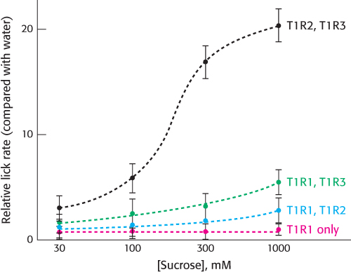
Most sweet compounds are carbohydrates, energy rich and easily digestible. Some noncarbohydrate compounds such as saccharin and aspartame also taste sweet. Members of a second family of 7TM receptors are expressed in taste-receptor cells sensitive to sweetness. The three members of this family, referred to as T1R1, T1R2, and T1R3, are distinguished by their large extracellular domains compared with those of the bitter receptors. Studies in knockout mice have revealed that T1R2 and T1R3 are expressed simultaneously in mice able to taste carbohydrates (Figure 33.16). Thus, T1R2 and T1R3 appear to form a specific heterodimeric receptor responsible for mediating the response to sugars. This heterodimeric receptor also responds to artificial sweeteners and to sweet-tasting proteins and therefore appears to be the receptor responsible for responses to all sweet tastants. Note that T1R2 and T1R3 do respond to sweet tastants individually but only at very high concentrations of tastant.

969

The requirement for an oligomeric 7TM receptor for a fully functional response is surprising, considering our previous understanding of 7TM receptors. This discovery has at least two possible explanations. First, the sweet receptor could be a member of a small subset of the 7TM-receptor family that functions well only as oligomers. Alternatively, many 7TM receptors may function as oligomers, but this notion is not clear, because these oligomers contain only one type of 7TM-receptor subunit. Further studies will be required to determine which of these explanations is correct.

Umami, the taste of glutamate and aspartate, is mediated by a heterodimeric receptor related to the sweet receptor

The family of receptors responsible for detecting sweetness is also responsible for detecting amino acids. In human beings, only glutamate and aspartate elicit a taste response. Studies similar to those for the sweet receptor revealed that the umami receptor consists of T1R1 and T1R3. Thus, this receptor has one subunit (T1R3) in common with the sweet receptor but has an additional subunit (T1R1) that does not participate in the sweet response. This conclusion is supported by the observation that mice in which the gene for T1R1 is disrupted do not respond to aspartate but do respond normally to sweet tastants; mice having disrupted genes for both T1R1 and T1R3 respond poorly to both umami and sweet tastants.

Salty tastes are detected primarily by the passage of sodium ions through channels

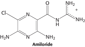
Salty tastants are not detected by 7TM receptors. Rather, they are detected directly by their passage through ion channels expressed on the surface of cells in the tongue. Evidence for the role of these ion channels comes from examining known properties of Na+ channels characterized in other biological contexts. One class of channels, characterized first for its role in salt reabsorption, is thought to be important in the detection of salty tastes because these channels are sensitive to the compound amiloride, which mutes the taste of salt and significantly lowers sensory-neuron activation in response to sodium.

An amiloride-sensitive Na+ channel comprises four subunits that may be either identical or distinct but in any case are homologous. An individual subunit ranges in length from 500 to 1000 amino acids and includes two presumed membrane-spanning helices as well as a large extracellular domain in between them (Figure 33.17). The extracellular region includes two (or, sometimes, three) distinct regions rich in cysteine residues (and, presumably, disulfide bonds). A region just ahead of the second membrane-spanning helix appears to form part of the pore in a manner analogous to that of the structurally characterized potassium channel. The members of the amiloride-sensitive Na+ -channel family are numerous and diverse in their biological roles. We shall encounter them again in the context of the sense of touch.

Figure 33.17: Schematic structure of the amiloride-sensitive sodium channel. Only one of the four subunits that constitute the functional channel is illustrated. The amiloride-sensitive sodium channel belongs to a superfamily having common structural features, including two hydrophobic membrane-spanning regions, intracellular amino and carboxyl termini, and a large, extracellular region with conserved cysteine-rich domains.

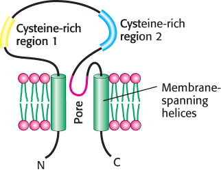
Sodium ions passing through these channels produce a significant transmembrane current. Amiloride blocks this current, accounting for its effect on taste. However, about 20% of the response to sodium remains even in the presence of amiloride, suggesting that other ion channels also contribute to salt detection.

Sour tastes arise from the effects of hydrogen ions (acids) on channels

Like salty tastes, sour tastes are detected by direct interactions with ion channels, but the incoming ions are hydrogen ions (in high concentrations) rather than sodium ions. For example, in the absence of high concentrations of sodium, hydrogen ion flow can induce substantial transmembrane currents through amiloride-sensitive Na + channels. However, hydrogen ions are also sensed by mechanisms other than their direct passage through membranes. Binding by hydrogen ions blocks some potassium ion channels and activates other types of channels. Together, these mechanisms lead to changes in membrane polarization in sensory neurons that produce the sensation of sour taste. We shall consider an additional receptor related to taste, one responsible for the “hot” taste of spicy food, when we examine mechanisms of touch perception.

970