Chapter 27. The Visual Pathway

Introduction

axon
part of a neuron specialized to send impulses to other neurons
bipolar cells
the middle layer of cells within the retina, connecting photoreceptors to ganglion cells
cones
photoreceptor cells in the eye specialized for detailed color vision in bright light
cornea
clear outer surface of the eyeball
ganglion cells
top layer of cells within the retina; axons of these cells form the optic nerve
iris
ring of muscle in the eye that controls the size of the pupil
lens
structure inside the eye that changes shape to focus images on the retina
neural impulse
an electrical signal that carries a message along an axon
occipital lobes
part of the cerebral cortex located in the lower back portion of the brain; contains the visual cortex
optic chiasm
X-shaped structure where the two optic nerves merge to combine and sort the information from the two eyes
optic nerve
contains the axons of the ganglion cells; carries visual information from the eyes toward the brain
photoreceptors
special cells in the retina that detect light
pupil
adjustable opening that allows light to enter the eye
retina
light-sensitive inner surface of the eye, containing rods and cones
rods
photoreceptor cells in the eye specialized for monochrome vision in dim light
thalamus
main relay center in the brain for sensory information on the way to processing areas in the cortex
visual cortex
area in the occipital lobes that receives and processes information from the eyes
The Visual Pathway
true
true
true
The image is a picture of the visual system inside the head. The eyes are attched to the visual cortex on both sides of the back of the brain. Between the eyes and the visual cortex is round element called the thalamus.
Learning Objectives:

Name the major structures of the visual system.

Understand the role that each structure plays in transmitting visual information.

Review

concept_review

Review

Select the NEXT button to continue with the Review.

The image is a horizontal cross-section of the brain so that we are looking down from the top of the brain into the various components of the visual system.  Two optic nerves leave each eye, one carrying information from the left side of the eye, another carrying information from the right side of the eye. The optic nerves combine at the optic chiasm. At this point the optic nerve from the left side of each eye come together to create an optic tract that goes to the left side of the brain.  The optic nerve from the right side of each eye come together to create an optic tract that goes to the right side of the brain. The optic tracts travel to the lateral geniculate nucleus of the thalamus on their respective sides of the brain. Then the optic tracts transition through optic radiation to the primary visual cortex on each side of the brain.

1. The visual pathway consists of all the structures in the visual system, from the eyes at the front of the head to the visual cortex, located in the brain’s occipital lobes at the back of the head. Let’s take a quick tour of this pathway.

Review

concept_review

Review

Select the NEXT button to continue with the Review.

The structures of the eye are labeled from front to back of the eye as follows: cornea, iris, pupil, lens, and retina. The cornea is in the front of the eye.  The iris and pupil are located directly behind the cornea. The lens is at the front of the eye, behind the pupil. The retina is the inner lining of the eye ball. An arrow points from outside the eye through the elements of the eye to a spot on the back of the retina.  This arrow is labeled as 'light'.

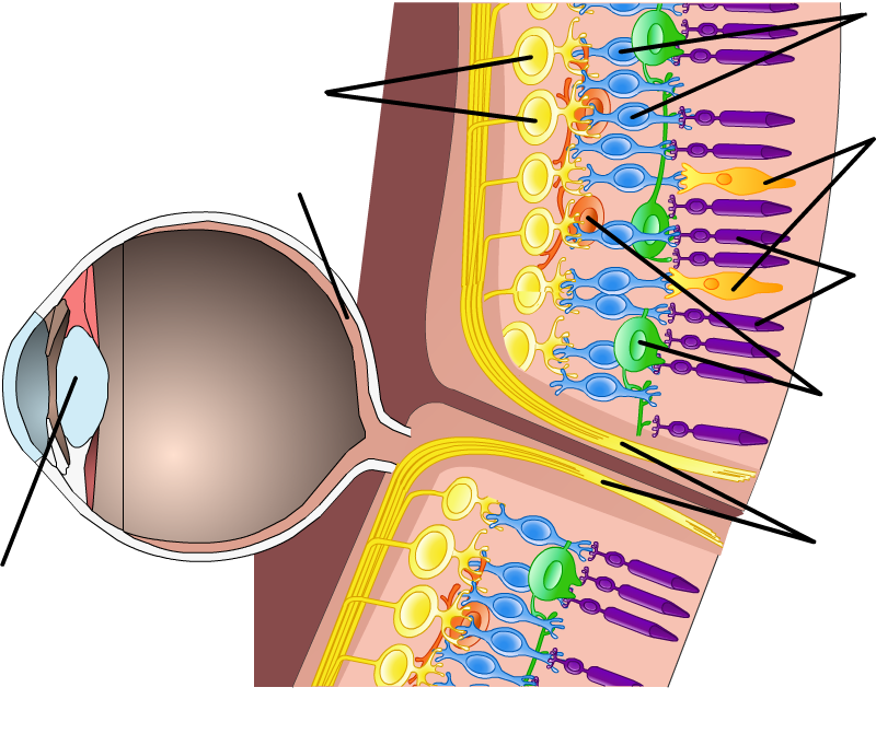
2. Light from the outside world passes through the cornea and then through the pupil, the opening in the ring of muscle called the iris. The lens focuses the light on the retina, where the process of seeing actually begins.

Review

concept_review

Review

Select the NEXT button to continue with the Review.

The structure of the eye has a small square section of the retina cut out and enlarged showing the different layers of cells in this part of the eye. The top layer of cells are labeled as ganglion cells. The bottom layer of cells are labeled as rods and cones.

3. The retina contains two types of photoreceptors: rods specialized for monochrome vision in dim light, and cones specialized for color vision in bright light. The photoreceptors capture the light energy and convert it to neural impulses. These signals are passed through the bipolar cells to the ganglion cells, whose axons form the optic nerve.

Review

concept_review

Review

Select the NEXT button to continue with the Review.

The image is a horitzontal cross-section of the visual system. Two optic nerves leave each eye, one carrying information from the left side of the eye, another carrying information from the right side of the eye. The optic nerves combine at the optic chiasm. At this point the optic nerve from the left side of each eye come together to create an optic tract that goes to the left hemisphere of the brain. The optic nerve from the right side of each eye come together to create an optic tract that goes to the right hemipshere of the brain.

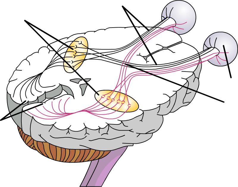
4. The visual information leaves the eye on the optic nerve and travels to the optic chiasm, where the signals from the portion of the retina nearest the nose cross to the other side of the brain.

Review

concept_review

Review

Select the NEXT button to continue with the Review.

The image is a horizontal cross-section of the brain so that we are looking down from the top of the brain into the various components of the visual system. Two optic nerves leave each eye, one carrying information from the left side of the eye, another carrying information from the right side of the eye. The optic nerves combine at the optic chiasm.  At this point the optic nerve from the left side of each eye come together to create an optic tract that goes to the left side of the brain.  The optic nerve from the right side of each eye come together to create an optic tract that goes to the right side of the brain.  The optic tracts travel to the lateral geniculate nucleus of the thalamus on their respective sides of the brain. Then the optic tracts transition through optic radiation to the primary visual cortex on each side of the brain.

5. The optic chiasm separates the visual information from each side of the retina. The visual area of the thalamus then relays the information to the visual cortex, which processes the information and constructs a representation of the visual world. At this point, the person “sees” the light.

Practice 1: Light Enters the Eye

video
true

Practice 1: Light Enters the Eye

Select the PLAY button to view the first stage of the visual pathway.

asset/activities/visual_pathway/video/anim1.mp4

Practice 2: Events in the Retina

video
true

Practice 2: Events in the Retina

Select the PLAY button to view the second stage of the visual pathway.

asset/activities/visual_pathway/video/anim2.mp4

Practice 3: Impulses Travel to the Brain

video
true

Practice 3: Impulses Travel to the Brain

Select the PLAY button to view the last stage of the visual pathway, as the impulses travel to the brain.

asset/activities/visual_pathway/video/anim3.mp4

Quiz 1

dnd_test

Quiz 1

Drag each label to the line pointing to the appropriate structure. When all the labels have been placed, select the CHECK ANSWER button.

Select the NEXT button and move to Quiz 2.
Perhaps you should go back to review the structures of the eye involved in the visual pathway.
Cones
Ganglion cells
Retina
Rods
Connecting cells
Lens
Bipolar cells
Optic nerve

Quiz 2

dnd_test

Quiz 2

Drag each label to the line pointing to the appropriate structure. When all the labels have been placed, select the CHECK ANSWER button.

Select the NEXT button and move to Quiz 3.
Perhaps you should go back to review the structures of the brain involved in the visual pathway.
Optic chiasm
Retina
Thalamus
Optic nerve
Visual cortex

Quiz 3

matching_test

Quiz 3

Match the terms for visual pathway structures with their functions by dragging each colored circle to the appropriate gray circle. When all the circles have been placed, select the CHECK ANSWER button.

Select the NEXT button and move to the Conclusion.
Perhaps you should go back to review the structures involved in the visual pathway.
retina
thalamus
optic nerve
visual cortex
photoreceptors
ganglion cells
bipolar cells
lens
relay station between the eyes and the visual cortex
their axons form the optic nerve
change the light energy into neural impulses
changes shape to focus images on the retina
carries neural impulses from the retina toward the brain
area in the occipital lobe that receives and processes information from the eyes
light-sensitive inner surface of eye, containing the rods and cones
pass on the impulses from rods and cones

Conclusion

end_slide
The image is a picture of the visual system inside the head. The eyes are attached to the visual cortex on both sides of the back of the brain. Between the eyes and the visual cortex is round element called the thalamus.
Congratulations!
You have completed the activity Title