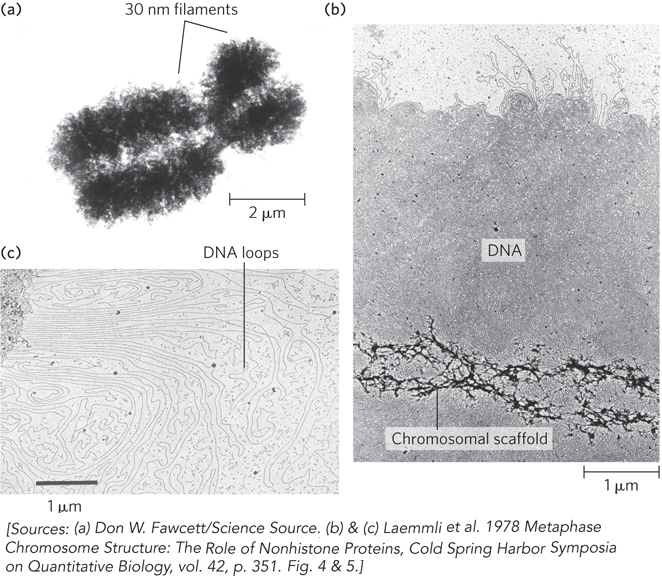
Loops of DNA attached to a chromosomal scaffold. (a) A swollen chromosome, produced in a buffer of low ionic strength, as seen in the electron microscope. Notice the appearance of 30 nm filaments (chromatin loops) at the margins. (b) Extraction of the histones leaves a proteinaceous chromosomal scaffold surrounded by naked DNA. (c) The DNA appears to be organized in loops attached at their base to the scaffold in the upper left corner. Note the different magnifications for the three images.