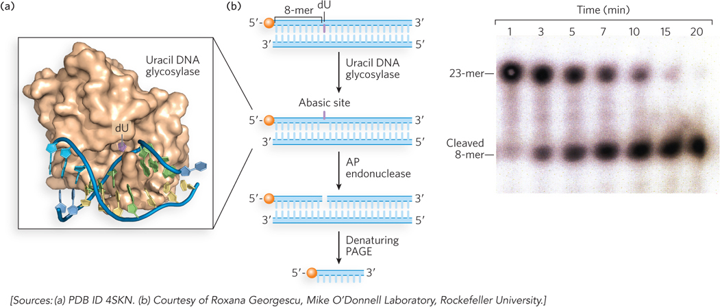
Uracil DNA glycosylase. (a) Human uracil DNA glycosylase (brown) bound to its DNA substrate. The uracil (purple) is flipped out of the DNA duplex and fits into the enzyme active site. (b) In this experiment, the DNA duplex, labeled at the 5′ end with 32P (orange balls), is treated with E. coli uracil DNA glycosylase for different time intervals to produce the abasic site, then with AP endonuclease to cleave the phosphodiester backbone. Electrophoresis in a denaturing polyacrylamide gel (PAGE), followed by autoradiography (right), shows results for the various time periods.