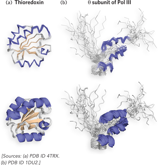
The structure of two proteins as determined by NMR. (a) Human thioredoxin (Mr 12,000). Multiple lines represent structures consistent with the restraints from the NMR data. One line is shown thicker than the rest to show the secondary elements within the structure. (b) The θ (theta) subunit (Mr 8,600) of DNA polymerase III (Pol III). The divergent models reflect the lack of restraints in disordered areas. The protein contains a region that lacked sufficient restraints to arrive at a unique solution and probably signifies a region of disordered residues.