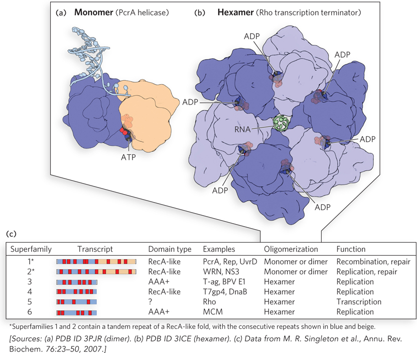
DNA helicases. (a) Enzymes of helicase superfamilies 1 and 2 (SF1 and SF2) have a conserved core structure, the RecA-like fold. Here, an ATP analog (black and red) that mimics ATP structure but is not hydrolyzed is bound at the interface of the two domains in a monomeric helicase PcrA. The use of such ATP analogs is often necessary to obtain a structure that illustrates the complex. (b) The core of helicases of SF3 through SF6 consists of six subunits with nucleotide-binding pockets at the subunit-subunit interfaces. Shown here is the Rho transcription termination factor (discussed in Chapter 15). In the structure shown, ADP was present in the crystallization mixture, and its bound location in the resulting structure indicates the ATPase active site. (c) Helicase superfamilies. Core domains (blue and beige) and positions of the conserved sequence motifs that characterize a protein as a helicase (red) are shown for each family (“Transcript”). The sequence motifs are universal structural elements in all helicases and include motifs involved in the binding and hydrolysis of a nucleoside triphosphate. There are two different ATPase domain types in helicases, as shown in the “Domain type” column. Well-studied examples of each family, typical oligomeric structure, and functions are listed in the remaining three columns.