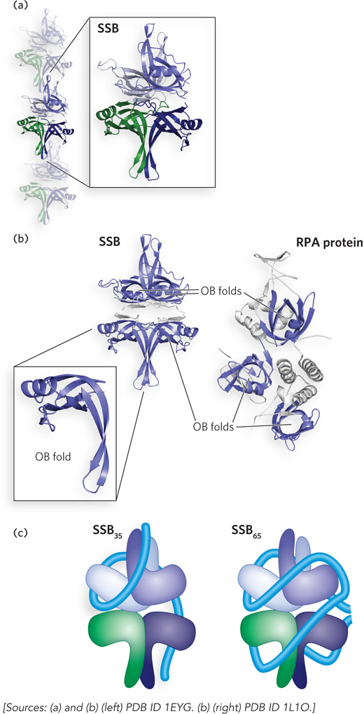
Binding of single-stranded DNA to single-stranded DNA–binding proteins. (a) Single-stranded DNA–binding protein (SSB) can bind in a filamentous form on single-stranded DNA. A single SSB tetramer is highlighted. (b) The key structural element that functions in single-stranded DNA binding is an OB fold. The basic fold consists of two three-stranded antiparallel β-sheets that share strand 1, with the strands ordered 1-2-3-5-4-1 (with the numbering coinciding to their order in the linear protein sequence). The sheets are tightly curved to form a small β-barrel, and an α-helix often connects strands 3 and 4. The structure can vary in appearance due to differences in lengths of the β-strands. The four OB folds in an SSB tetramer are highlighted in blue (left). The eukaryotic replication protein A (RPA) binds to single-stranded DNA in a similar fashion, using the OB folds in each subunit of the heterotrimer (right). (c) SSB can bind to single-stranded DNA in multiple binding modes, with the two most prominent modes shown in this schematic. The blue tube represents bound single-stranded DNA.