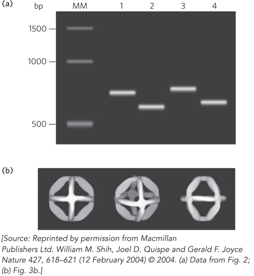
FIGURE 2 Gel electrophoresis and electron microscopy revealed the DNA octahedron assembly through base pairing. (a) Agarose gel electrophoresis of octahedron-forming DNA under different conditions. Lane MM: molecular marker lane with DNA size standards (number of base pairs (bp) is indicated on the left). Lane 1: 1,669-nucleotide strand (heavy chain) folded in the absence of Mg2+. Lane 2: 1,669-nucleotide strand folded in the presence of Mg2+. Lane 3: 1,669-nucleotide strand and 40-nucleotide light chains folded in the absence of Mg2+. Lane 4: 1,669-nucleotide strand and 40-nucleotide light chains folded in the presence of Mg2+. The Mg2+ shields the negative charges of the backbone phosphates and also promotes base pairing, creating a more compact structure that migrates more quickly through the gel. The resolution of the gel is not sufficient to detect differences in mobility of the DNA with or without the 40-nucleotide light chains. (b) Three views of the three-dimensional images of assembled DNA octahedra generated computationally, using electron micrographs as the starting point.