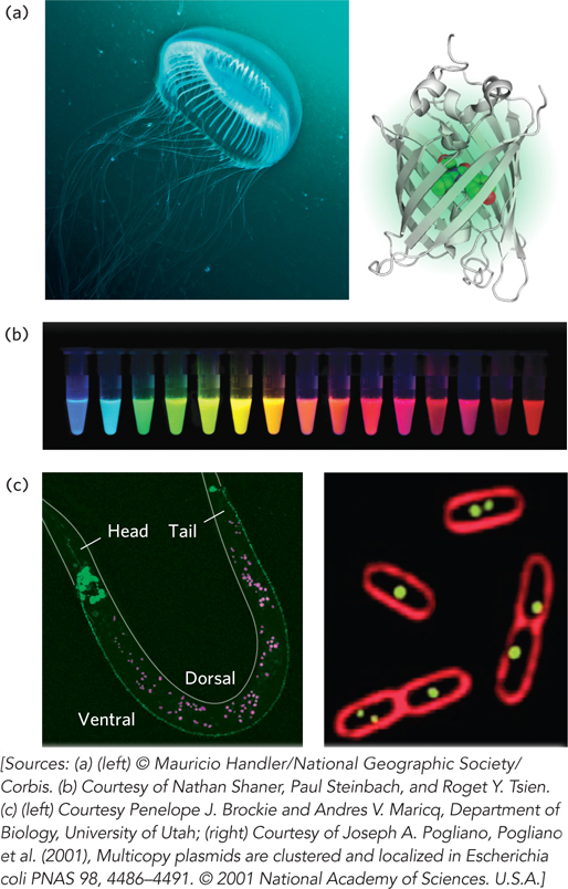
Green fluorescent protein (GFP). (a) The source of GFP is the jellyfish Aequorea victoria (left); the bioluminescent photo-organs are visible. The GFP protein has a β-barrel structure (right); the fluorophore (red) is in the center of the barrel. (b) Variants of GFP are now available in almost any color of the visible spectrum. (c) Caenorhabditis elegans, a nematode worm (left), containing a GLR1-GFP fusion protein. GLR1 is a glutamate receptor of nervous tissue. In the E. coli cells (right), the membranes are stained with a red fluorescent dye. The cells are expressing a protein that binds to a resident plasmid, fused to GFP. The green spots indicate the locations of plasmids.