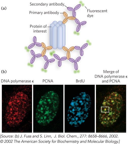
Indirect immunofluorescence. (a) The protein of interest is bound to a primary antibody, and a secondary antibody is added; this secondary antibody, with one or more attached fluorescent groups, binds to the primary antibody. Multiple secondary antibodies can bind the primary antibody, amplifying the signal. If the protein of interest is in the interior of the cell, the cell is fixed and permeabilized, and the two antibodies are added in succession. (b) The end result is an image in which bright spots indicate the location of the protein of interest in the cell. The images here show a nucleus from a human fibroblast, stained with antibodies and fluorescent labels for DNA polymerase ε, for PCNA, an important polymerase accessory protein, and for bromo-