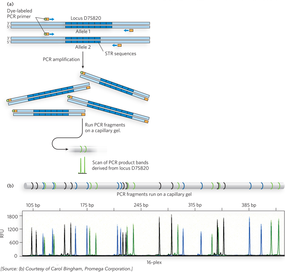
FIGURE 1 (a) STR loci can be analyzed by PCR. Suitable PCR primers (with an attached dye to aid in subsequent detection) are targeted to sequences on each side of the STR, and the region between them is amplified. If the STR sequences have different lengths on the two chromosomes of an individual’s chromosome pair, two PCR products of different lengths result. (b) The PCR products from amplification of up to 16 STR loci can be run on a single capillary acrylamide gel (a “16- s— R—