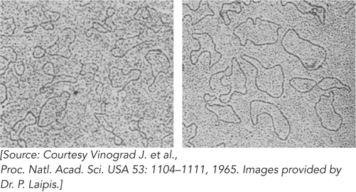
FIGURE 1
These electron micrographs of polyoma virus DNA, form I (20S) at left and form II (16S) at right, show the unexpected circular patterns.