9.2 DNA SUPERCOILING
Cellular DNA, as we have seen, is a very long molecule that must somehow be made to fit inside the cell, implying a high degree of structural organization. The folding mechanism must not only pack the DNA but permit access to the information in the DNA in processes such as replication and transcription. Any consideration of how this is accomplished requires an understanding of an important property of DNA structure known as supercoiling.
Supercoiling simply means the coiling of a coil. An old-fashioned telephone cord, for example, is typically a coiled wire. If one end of the cord is manually twisted, the coiled cord forms supercoils (Figure 9-6). DNA is coiled in the form of a double helix, with both strands coiling around an axis. DNA supercoiling is the further coiling of that axis upon itself (Figure 9-7). As described below, supercoiled DNA (sometimes called superhelical DNA) is generally a manifestation of structural strain. The DNA can be overwound to generate supercoils. However, underwinding of the DNA also imparts strain and results in supercoils. When there is no net coiling of the DNA axis upon itself, the molecule is referred to as relaxed DNA (see the How We Know section at the end of this chapter). Supercoiling occurs in all chromosomal DNAs in all cells, as well as in viruses that have a double-stranded DNA genome or generate double-stranded DNA as a replication intermediate.
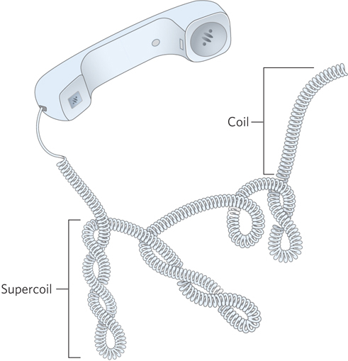
Figure 9-6: Supercoils. An old-fashioned phone cord is coiled like a DNA helix, and the coiled cord can supercoil.
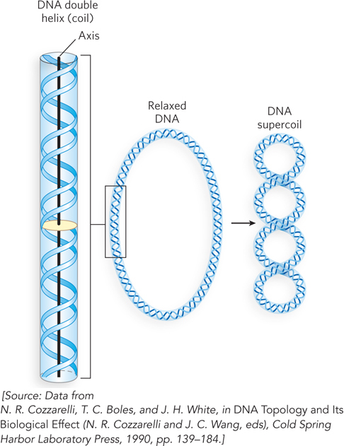
Figure 9-7: DNA supercoiling. When the DNA double helix is coiled on itself, it forms a new helix, or superhelix. The DNA superhelix is usually referred to as a supercoil.
It would be difficult to compact DNA without some form of supercoiling. Perhaps less evident is that replication and transcription of DNA also affect, and are affected by, supercoiling. Both processes require a separation of DNA strands, which is complicated by the helical interwinding of the strands (Figure 9-8). As discussed in Section 9.3, specialized enzymes found in all organisms alleviate the stress of replication and transcription by introducing or relaxing supercoils.
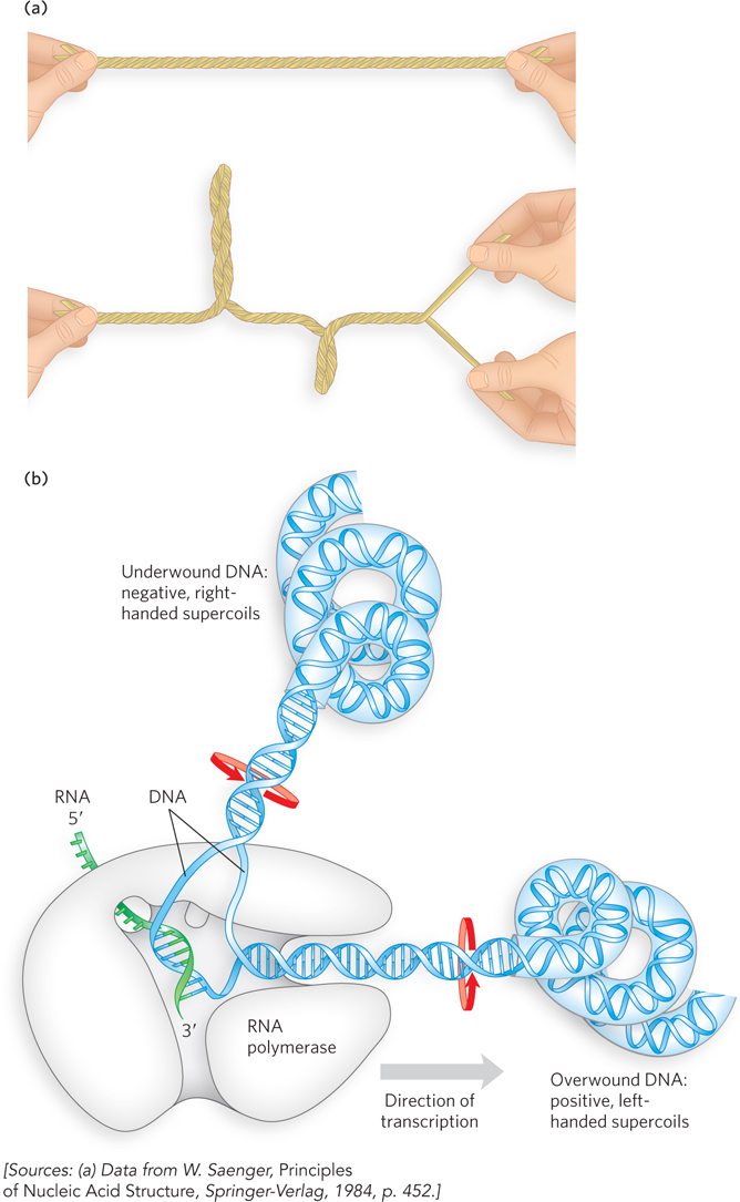
Figure 9-8: The effects of replication and transcription on DNA supercoiling. Because DNA is a double-helical structure, strand separation leads to added stress and supercoiling if the DNA is constrained (not free to rotate) ahead of the strand separation. (a) The general effect can be illustrated by twisting two strands of a rubber band about each other to form a double helix. If one end is constrained, separating the two strands at the other end will lead to twisting. (b) In a DNA molecule, the progress of a DNA polymerase or RNA polymerase (as shown) along the DNA involves separation of the strands. Chromosomal DNAs are typically complexed with many proteins that inhibit free rotation. As the polymerase moves on a DNA molecule that is rotationally constrained, the DNA becomes overwound ahead of the enzyme (upstream; positive supercoils) and underwound behind it (downstream; negative supercoils). Red arrows indicate the direction of winding.
Inside the cell, a DNA molecule must bend, and it inevitably becomes supercoiled as its axis twists on itself. However, even when extracted and purified, many circular DNA molecules remain highly supercoiled, even in the absence of protein and other cellular components. This indicates that supercoiling is an intrinsic property of DNA tertiary structure, as opposed to an incidental result of spatial constriction. Supercoiling is the direct result of structural strain caused by the active underwinding of the DNA—that is, the removal of helical turns relative to the most stable structure of B-form DNA. DNA underwinding is catalyzed by enzymes called topoisomerases, and the degree of DNA underwinding is highly regulated in every cell.
Several measurable properties of supercoiling have been established, and the study of supercoiling has provided many insights into DNA structure and function. This work has drawn heavily on concepts derived from topology, a branch of mathematics that studies the properties of an object that do not change under continuous deformations (deformations that do not involve breaking the DNA). In the context of DNA topology, continuous deformations include conformational changes due to stretching, thermal motion, or interaction with proteins or other molecules. The twisting experiments in the Bustamante laboratory, described in this chapter’s Moment of Discovery, provide an example of continuous deformation. Discontinuous deformations involve DNA strand breakage. Topological properties of DNA can be changed only by the breakage and rejoining (religation) of the backbone of one or both DNA strands.
Most Cellular DNA Is Underwound
The simplest examples of supercoiling are provided by small circular DNAs. These might include plasmids and small, double-stranded viral DNAs. When these DNA molecules have no breaks in either strand, they are referred to as closed-circular DNAs. If the double helix of a closed-circular molecule has the features of B-form structure (see Figure 6-17), with one turn of the double helix per 10.5 bp, it is relaxed rather than supercoiled (Figure 9-9). Supercoiling results when DNA is subject to some form of structural strain. However, purified closed-circular DNA is rarely relaxed, regardless of its biological origin. Furthermore, all DNA derived from a given cellular source has a characteristic degree of supercoiling. DNA structure is therefore maintained with a degree of strain that is regulated by the cell and, in turn, induces the observed supercoiling.
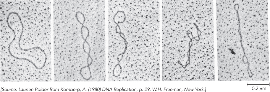
Figure 9-9: Relaxed and supercoiled closed-circular plasmid DNAs. The scanning electron micrograph on the far left shows relaxed DNA. Increased supercoiling is shown, from left to right.
In almost every instance in nature, the strain is a result of underwinding of the DNA double helix in the closed circle. In DNA underwinding, the molecule has fewer helical turns than would be expected for the B-form structure. Consider, for example, an 84 bp segment of a circular DNA in the relaxed state: it would contain eight double-helical turns, one for every 10.5 bp (Figure 9-10a). If one of these turns were removed, there would be (84 bp)/7 = 12.0 bp per turn, rather than the 10.5 found in B-DNA (Figure 9-10b). This is a deviation from the most stable DNA form, and the molecule would be thermodynamically strained as a result. Generally, much of this strain would be accommodated by coiling the axis on itself, forming a supercoil (Figure 9-10c). Some of the strain in this 84 bp segment would simply become dispersed in the untwisted structure of the larger DNA molecule. In principle, the strain induced by this degree of net DNA underwinding could also be accommodated by separating the two DNA strands over a distance of about 10 bp (Figure 9-10d). In isolated closed-circular DNA, strain introduced by underwinding is generally accommodated by supercoiling rather than strand separation, because coiling the axis of the DNA usually requires less energy than breaking the hydrogen bonds that stabilize paired bases. Note, however, that the underwinding of DNA in vivo eases the task of DNA strand separation by enzymes such as DNA and RNA polymerases, and thus facilitates access to the information in the DNA.
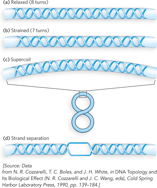
Figure 9-10: The effects of DNA underwinding.
Every cell actively underwinds its DNA with the aid of enzymes (see Section 9.3), and the resulting strained state represents a form of stored energy. Underwinding thus accomplishes two things. First, cells maintain DNA in an underwound state in part to promote its compaction by coiling. Second, underwinding facilitates strand separation and enzymatic access to the encoded information; underwinding is thus important to the enzymes of DNA replication and transcription, which must bring about strand separation as part of their function. In this way, two biological problems related to DNA—compaction and information access—are addressed at once by active DNA underwinding.
The underwound state can be maintained only if the DNA is a closed circle or, if linear, is bound and stabilized by proteins so that the strands are not free to rotate about each other. If there is a break in one strand of an isolated, protein-free circular DNA, free rotation at that point will cause the underwound DNA to revert spontaneously to the relaxed state. In a closed-circular DNA molecule, however, the number of helical turns cannot be changed without at least transiently breaking one of the DNA strands. The number of helical turns in a DNA molecule therefore provides a precise description of supercoiling. In the linear chromosomes of eukaryotic cells, DNA underwinding is maintained by bound proteins that constrain the DNA in an elaborate structure called chromatin. In chromatin, large loops of DNA are constrained at their base, such that each loop is topologically fixed as if it were circular (Figure 9-11; we discuss this further in Chapter 10).
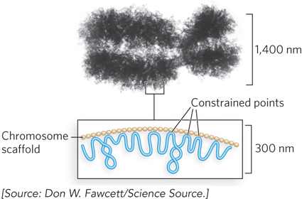
Figure 9-11: Loops in a eukaryotic chromosome constrained by scaffold proteins. The chromatin scaffold is attached to the chromosome at intervals, with the DNA between the attachment points defining loops that are topologically constrained.
DNA Underwinding Is Defined by the Topological Linking Number
The linking number (Lk) is a topological property of double-stranded DNA—that is, it does not change when the DNA is bent or deformed. To define linking number, imagine the separation of the two strands of a double-stranded circular DNA. If the two circular strands are interlocked as shown in Figure 9-12a, they are effectively linked by what we define as a topological bond. Even if all hydrogen bonds and base-stacking interactions were removed so that the strands were not in physical contact, the two strands would still be interlocked like the links of a chain link fence. This is the topological linkage. If we think of one of the circular strands as the boundary of a surface (such as the soap film on the loop of a bubble wand before you blow a bubble), the linking number can be defined as the number of times the second strand pierces this surface (Figure 9-12b). The linking number for a closed-circular DNA is always an integer. By convention, if the DNA strands are interwound in a right-handed helix, the linking number is positive (+); for strands interwound in a left-handed helix, the linking number is negative (−). Negative linking numbers are, for all practical purposes, not encountered in DNA.
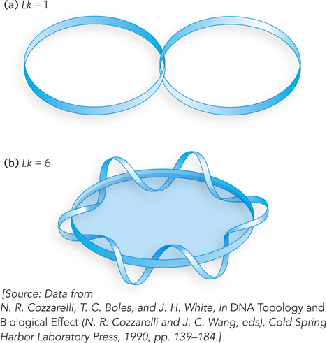
Figure 9-12: Linking number (Lk). Each blue ribbon represents one strand of a double-stranded DNA molecule. (a) The linking number of the molecule is 1. (b) The linking number is 6. One of the strands is kept untwisted for illustrative purposes, to define the border of an imaginary surface (solid blue oval). The number of times the twisting strand (its relative length exaggerated here) penetrates this surface is the linking number.
Consider a closed-circular DNA with 2,100 bp (Figure 9-13a). When the molecule is relaxed, the linking number is equal to the number of base pairs divided by the number of base pairs per turn, 10.5; in this case, Lk = 200. In unstrained B-form DNA, this is the number of times that one strand would pierce the imaginary surface defined by the other strand (see Figure 9-12). DNA can have a topological property such as a linking number only if both strands are intact. If there is a break in either strand, the strands can, in principle, be unraveled and separated; in this case, no topological bond exists and Lk is undefined (Figure 9-13b).
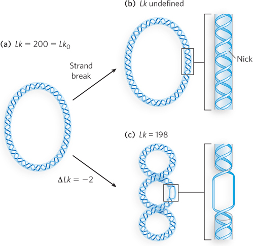
Figure 9-13: Linking number of closed-circular DNAs. A 2,100 bp molecule is shown in three forms: (a) relaxed, Lk = 200; (b) relaxed with a nick in one strand, Lk undefined; (c) underwound by two turns, Lk = 198. The underwound molecule is generally supercoiled, but underwinding also facilitates the separation of DNA strands.
We can now describe DNA underwinding in terms of changes in the linking number (ΔLk). The linking number in relaxed DNA, Lk0, is used as a reference. For the molecule in Figure 9-13a, Lk0 = 200. If the linking number changes, such that Lk = 198 (Figure 9-13c), the change (number of turns removed) can be described by the equation:
In our example, ΔLk = 198 − 200 = −2.
We can also express the change in linking number independent of the length of the DNA molecule. This quantity, usually called the superhelical density (σ), is a measure of the number of turns removed relative to the number present in relaxed DNA:
In the Figure 9-13c example, σ = −0.01, which means that 1% (2 of 200) of the helical turns present in the DNA (when it is relaxed, in its B form) have been removed. The degree of underwinding in cellular DNAs generally falls in the range of 5% to 7%; that is, σ = −0.05 to −0.07. The negative sign indicates that the change in the linking number is due to underwinding of the DNA. The supercoiling induced by underwinding is therefore defined as negative supercoiling. Conversely, under some conditions DNA can be overwound, resulting in positive supercoiling.
Notice that negative supercoiling results in a twisting of the axis of the DNA to form a right-handed superhelix, and positive supercoiling results in a left-handed superhelix (Figure 9-14). Supercoiling is not a random process; it is largely prescribed by the torsional strain imparted to the DNA by decreasing or increasing the linking number relative to that of B-DNA.
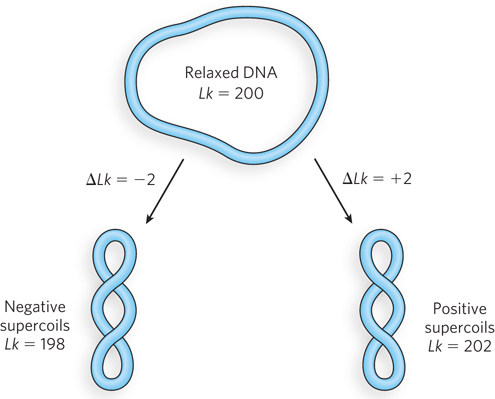
Figure 9-14: Negative and positive supercoils. For the relaxed DNA in Figure 9-13a, underwinding or overwinding by two helical turns (ΔLk = ±2) produces negative or positive supercoiling, respectively. Notice that the DNA axis in the two forms twists in opposite directions.
The linking number can be changed by ±1 by breaking one DNA strand, rotating one of the ends 360° about the unbroken strand, and rejoining the broken ends. This reaction is catalyzed by topoisomerases (see Section 9.3). The change in linking number has no effect on the number of base pairs or the number of atoms in the circular DNA molecule. Two forms of a circular DNA that differ only in a topological property such as linking number are referred to as topoisomers.
We can break down the linking number into two structural components, writhe (Wr) and twist (Tw) (Figure 9-15). Writhe (Wr) is a measure of the coiling of the helical axis, and twist (Tw) describes the local twisting or spatial relationship of neighboring base pairs. When the linking number changes, some of the resulting strain is usually compensated for by writhe (supercoiling) and some by changes in twist, giving rise to the equation:
Tw and Wr need not be integers. Twist and writhe are geometric rather than topological properties, because they may be changed by deformation of a closed-circular DNA molecule. Tw and Wr may change in a reciprocal manner without altering Lk. That is, Lk can remain unchanged when either Tw or Wr increases by a given amount and the other decreases by that same amount.
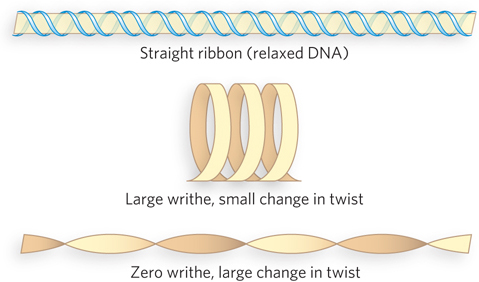
Figure 9-15: Writhe and twist. The beige ribbon represents the axis of a relaxed DNA molecule. Strain from underwinding of the DNA can manifest as writhe or twist. Topological changes in the linking number are usually accompanied by geometric changes in both writhe and twist.
In addition to causing supercoiling and making strand separation somewhat easier, the underwinding of DNA facilitates structural changes in the molecule. Although these changes are of less physiological importance, they help illustrate the effects of underwinding. Recall that a cruciform generally contains a few unpaired bases (see Figure 6-20b); DNA underwinding helps maintain the required strand separation in regions where palindromic sequences allow cruciform formation (Figure 9-16). Underwinding of a right-handed DNA helix also facilitates the formation of short stretches of left-handed Z-DNA in regions where the base sequence is consistent with the Z form (see Chapter 6).
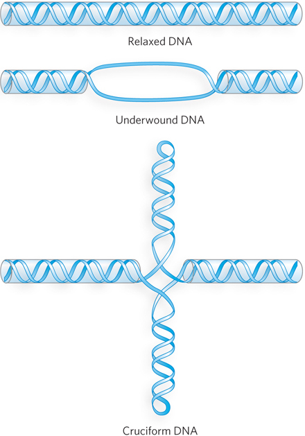
Figure 9-16: Promoting cruciform structures by DNA underwinding. Cruciforms can form at palindromic sequences, but they seldom occur in relaxed DNA because linear DNA accommodates more paired bases than the cruciform structure. DNA underwinding facilitates the partial strand separation required for promoting cruciform formation at appropriate sequences.
DNA Compaction Requires a Special Form of Supercoiling
All supercoiled DNA molecules are similar in several respects. In the laboratory, the supercoils are right-handed in a negatively supercoiled DNA molecule, such as a supercoiled plasmid isolated from a bacterium. The supercoiled plasmids tend to be extended and narrow rather than compacted, often with multiple branches (Figure 9-17a). At the superhelical densities normally encountered in cells, the length of the supercoil axis (the axis about which the supercoils turn), including branches, is about 40% of the length of the DNA. This type of supercoiling is referred to as plectonemic supercoiling (Figure 9-17b). This term can be applied to any structure with strands intertwined in some simple and regular way, and it is a good description of the general structure of supercoiled DNA in solution. In cells, it is found in smaller DNAs such as plasmids. Larger genomic DNAs are generally compacted in a different way.
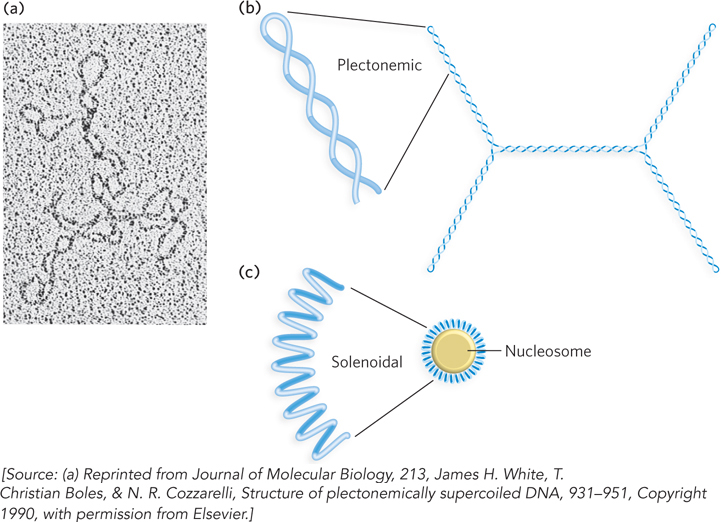
Figure 9-17: Plectonemic and solenoidal supercoiling. (a) Electron micrograph of plectonemically supercoiled plasmid DNA. (b) Plectonemic supercoiling consists of extended right-handed coils. (c) Solenoidal supercoiling of the same DNA molecule depicted in (b), drawn to scale. Solenoidal negative supercoiling consists of tight left-handed turns about an imaginary tubelike structure. Solenoidal supercoiling provides a much greater degree of compaction.
Plectonemic supercoiling does not produce sufficient compaction to package genomic DNA in the cell. A second form, solenoidal supercoiling, can be adopted by an underwound DNA in the cell (Figure 9-17c). Instead of the extended right-handed supercoils characteristic of the plectonemic form, solenoidal supercoiling involves tight left-handed turns, much like a garden hose neatly wrapped on a reel. Although their structures are very different, plectonemic and solenoidal supercoiling are two forms of negative supercoiling that can be adopted by the same segment of underwound DNA. The two forms are readily interconvertible. The plectonemic form is more stable in solution, but the solenoidal form can be stabilized by protein binding and provides a much greater degree of compaction. Solenoidal supercoils are formed when DNA is wrapped around the nucleosomes that make up eukaryotic chromatin (see Chapter 10). Similarly, in bacteria, the tight wrapping of DNA around a variety of DNA-binding proteins gives rise to solenoidal supercoils. Solenoidal supercoiling is the primary mechanism by which underwinding contributes to genomic DNA compaction.
SECTION 9.2 SUMMARY
Most cellular DNAs are supercoiled. Underwinding decreases the total number of helical turns in the DNA relative to the relaxed, B form. To maintain an underwound state, DNA must be either a closed circle or, if linear, bound to protein.
Underwinding is quantified by the linking number (Lk), a topological parameter that describes the number of times two DNA strands are intertwined.
Underwinding is measured in terms of σ, the superhelical density; σ = ΔLk/Lk0. For cellular DNAs, σ is typically −0.05 to −0.07, which means that about 5% to 7% of the helical turns in the DNA have been removed. DNA underwinding facilitates strand separation by enzymes of DNA metabolism.
Plectonemic supercoiling includes right-handed branches and is the most common type of supercoiling in isolated DNA. Solenoidal supercoiling, an alternative form that produces a greater degree of compaction, is characterized by tight left-handed turns that are stabilized by wrapping the DNA around proteins; this occurs in eukaryotic and bacterial chromosomes.


