22.2 TRANSLATIONAL CONTROL IN THE CYTOPLASM

Regulation at the level of translation assumes a much more prominent role in eukaryotes than in bacteria and is observed in a range of cellular situations. In contrast to the tight coupling of transcription and translation in bacteria, transcripts generated in a eukaryotic nucleus must be processed and transported to the cytoplasm before translation. This can impose a significant delay on the availability of a protein function. When a rapid increase in protein production is needed, a translationally repressed mRNA already in the cytoplasm can be activated for translation without delay.

Translational control is responsible for activating the expression of proteins necessary for cell fate decisions. Translational regulation may play an especially important role in controlling the expression of certain very long eukaryotic genes (a few are measured in millions of base pairs!), for which transcription and mRNA processing can require many hours. Some genes are regulated at both the transcriptional and translational stages, with the latter playing a role in fine-tuning of cellular protein levels. In some non-nucleated cells, such as reticulocytes (immature red blood cells), transcriptional control is unavailable and translational control of stored mRNAs becomes essential. Translational controls are also essential during development, when the regulated translation of pre-positioned mRNAs creates a local gradient of the protein product (see Section 22.5).

Eukaryotes have at least three main mechanisms for regulating translation. First, various initiation factors are subject to phosphorylation by protein kinases. The phosphorylated forms are often less active and generally depress translation in the cell. Second, some proteins bind directly to mRNA and act as translational repressors. Many bind at specific sites in the 3′UTR and interact with other initiation factors bound to the mRNA, or they interact with the 40S ribosomal subunit to prevent initiation. Third, binding proteins can disrupt the interaction between eIF4E and eIF4G (recall from Chapter 18 that interaction between these initiation factors is required for proper assembly of the ribosome-mRNA complex). Such binding proteins are present in eukaryotes from yeast to mammals, and the mammalian versions are known as 4E-BPs (eIF4E binding proteins). When cell growth is slow, these proteins limit translation by binding to the site on eIF4E that normally interacts with eIF4G. When cell growth resumes or increases in response to growth factors or other stimuli, the binding proteins are inactivated by protein kinase–dependent phosphorylation.

766

The variety of translational regulation mechanisms in eukaryotes provides flexibility, allowing focused repression of a few mRNAs or global regulation of all cellular translation. Here we explore several examples in which these mechanisms come into play.

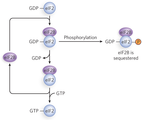
Initiation Can Be Suppressed by Phosphorylation of eIF2

Translation initiation in eukaryotes is a complex process involving multiple initiation factors that recruit ribosomes to mRNAs. Reversible phosphorylation of initiation factors plays a central role in regulating initiation. The phosphorylation is triggered by a wide range of cellular conditions, depending on cell type and function. One of the main phosphorylation pathways involves eIF2. In all eukaryotes, eIF2 is composed of three polypeptide subunits—eIF2α, eIF2β, and eIF2γ—that together bind the initiator tRNA and GTP. When Met-tRNAiMet binds to the peptidyl (P) site on the 40S ribosomal subunit, GTP is hydrolyzed, and eIF2-GDP dissociates from the initiator tRNA. Recycling of eIF2-GDP to eIF2-GTP requires eIF2B (Figure 22-6). If eIF2 is phosphorylated, eIF2B binding to eIF2 is nearly irreversible and the GDP is not dislodged. Because the cell has less eIF2B than eIF2, only a little phosphorylated eIF2 is needed to sequester all the eIF2B, thereby shutting down protein synthesis.

Figure 22-6: Halting the recycling of eIF2 by phosphorylation. The recycling of used eIF2 (eIF2-GDP), after it has served its role in translation initiation, is facilitated by a guanine nucleotide exchange factor, eIF2B. Phosphorylation of the eIF2 protein closes down this cycle, and thus controls translation rates, by tying up eIF2B.

This process has been well studied in mammalian reticulocytes. The maturation of reticulocytes into red blood cells includes destruction of the cell nucleus, leaving behind a hemoglobin-packed cell. Messenger RNAs deposited in the cytoplasm before loss of the nucleus allow for the replacement of hemoglobin. When reticulocytes become deficient in iron or heme, the translation of globin mRNAs is repressed. A protein kinase called HCR (hemin-controlled repressor) is activated, catalyzing phosphorylation of eIF2α, the smallest subunit of eIF2. When its eIF2α subunit is phosphorylated, eIF2 forms a stable complex with eIF2B, blocking dissociation of GDP after GTP hydrolysis and thus making these initiation factors unavailable for further rounds of translation. In this way, the reticulocyte coordinates the synthesis of globin with the availability of heme.

Phosphorylation of eIF2α regulates translation in other systems as well. For example, a double-stranded RNA–dependent protein kinase (PKR) phosphorylates eIF2α in some cells in response to viral infection. This helps block the translation of viral mRNAs and interferes with the viral life cycle. In yeast, activation of the kinase Gcn2 by nitrogen starvation leads to eIF2α phosphorylation and repression of most translation, until more nitrogen (and the amino acids that incorporate it) becomes available. In an interesting mechanistic twist, eIF2α phosphorylation in yeast induces translation of the transcription factor Gcn4 (which we discuss below).

The 3′UTR of Some mRNAs Controls Translational Efficiency

The 3′ untranslated region of an mRNA communicates with the 5′ end through protein-protein interactions between factors that bind specifically to the ends of a fully processed mRNA. This communication ensures that the mRNA is fully processed before translation begins. Circularization occurs when eIF4E bound at the 5′ terminus and poly(A) binding protein (PABP) bound at the 3′ poly(A) tail both interact with eIF4G (Figure 22-7a). Initiation of translation requires the recruitment of eIF4G by eIF4E, through a conserved motif in eIF4G that allows interaction with eIF4E. The same motif is used by other proteins to repress translation of certain mRNAs.

Figure 22-7: Functions of the 3′UTR and 5′UTR in translation initiation. (a) eIF4E bound to the 5′UTR, and poly(A) binding protein (PABP) bound to the 3′ poly(A) tail, are linked through their binding to eIF4G. The resulting looped complex facilitates recruitment of other translational regulators and ribosomal subunits for translation initiation. (b) For maternal mRNAs such as that derived from the Xenopus gene mos, eIF4E is initially sequestered by interaction with Maskin and CPE-binding protein (CPEB), with translation suppressed. Activation involves phosphorylation of CPEB, which in association with the cleavage and polyadenylation specificity factor (CPSF) promotes lengthening of the poly(A) tail and recruitment of PABP and eIF4G; eIFG displaces Maskin, allowing translation to start.

767

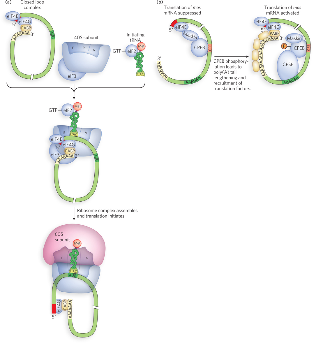
Regulation of translation by protein binding to the 3′UTR is especially important during the development of early embryos. Many maternal RNAs (deposited in the egg cytoplasm during oogenesis) have relatively short poly(A) tails (20 to 40 A residues), and their translation is suppressed until it is needed. Activation requires two sequences in the 3′UTR: the nuclear AAUAAA polyadenylation sequence and the cytoplasmic polyadenylation element (CPE, with consensus sequence UUUUUAUU). CPEs are bound by the protein CPEB (CPE-binding protein), which helps establish translational masking of maternal mRNAs by interacting with a protein known, appropriately, as Maskin. Maskin also interacts with eIF4E, functioning much like 4E-BPs in preventing binding of eIF4E to eIF4G. An example of this mechanism is the regulation of a maternal mRNA in Xenopus embryos, transcribed from a gene called mos (Figure 22-7b). To activate the mos mRNA, CPEB is first phosphorylated. This stimulates interaction between CPEB and the AAUAAA-binding protein CPSF (cleavage and polyadenylation specificity factor). In turn, CPSF recruits the cytoplasmic polyadenylation enzymes that lengthen the poly(A) tail on the mRNA. The longer poly(A) tails enable the binding of multiple copies of PABP. PABP recruits eIF4G, and eIF4G displaces Maskin and interacts with eIF4E. Translation then begins.

768

In addition to CPEBs, other types of proteins bind the 3′UTR of mRNA to regulate translation, including proteins of the PUF family (named for Pumilio and FBF, the first two proteins of this type to be discovered). PUF proteins are a highly conserved family of RNA-binding proteins associated with translational control in a variety of organisms, from yeast to mammals. PUF proteins typically include eight consecutive 40 residue repeat sequences, each of which contains characteristic aromatic and basic amino acids. The crescent-shaped structure of PUF proteins reveals two extended surfaces (Figure 22-8). Based on the location and effects of mutations, one surface probably binds to mRNA and the other to other regulatory proteins.

Figure 22-8: Structure and function of the PUF family protein Pumilio. The protein is crescent shaped, with 10 repeated α-helical segments, each consisting of about 40 amino acids. The 8 repeats in the middle (colored) are involved in mRNA binding. The 2 repeats on the ends (white) are important to Pumilio function, but do not directly participate in mRNA binding. Binding of a PUF family protein to the 3′UTR of an mRNA, as shown on the right, suppresses protein production either through interference with translation initiation by blocking assembly of translational factors or through promotion of mRNA degradation by recruiting RNA degradation enzymes. [Source: PDB ID 1M8Y.]

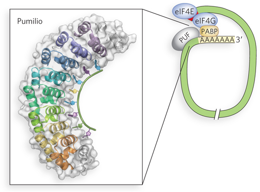
Each PUF protein is thought to regulate multiple mRNAs, because experiments have demonstrated the proteins’ ability to bind multiple targets. Researchers have engineered fruit flies to express a molecularly tagged version of Pumilio. The tagged protein was purified with its bound RNA partners, and the identities of the bound RNAs were determined using microarray technology (see Chapter 7). The study showed that many of the bound mRNAs shared a short, characteristic sequence in their 3′UTRs. Furthermore, the mRNAs tended to encode functionally related proteins, suggesting that Pumilio acts by binding to sets of mRNAs. Many of the proteins regulated by Pumilio function in developmental pathways considered in Section 22.5.

Once bound by the PUF protein, the targeted mRNAs are typically blocked from efficient translation on the ribosome. Although the mechanism of repression is not completely known, PUF proteins seem to block initiation. In some cases, PUF proteins may also increase the rate of mRNA degradation.

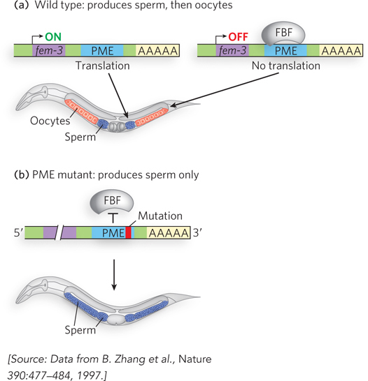
In the nematode Caenorhabditis elegans, the hermaphrodite produces sperm and oocytes, successively, during development of the germ line. This cell fate decision is controlled by the PUF family protein FBF (fem-3 binding factor), which binds to a site in the 3′UTR of the fem-3 gene called the PME (point mutation element) (see the How We Know section at the end of this chapter). This binding site was defined by gene mutants first isolated in the laboratory of Judith Kimble (see this chapter’s Moment of Discovery). When FBF is not present in the germ-line cells, translation of fem-3 transcripts proceeds and sperm cells are produced (Figure 22-9). When FBF protein is present, it binds the 3′UTR and blocks translation of fem-3 transcripts, and oocytes are produced.

Figure 22-9: The regulation of germ-line development in C. elegans hermaphrodites. (a) Hermaphrodites produce first sperm cells, then oocytes. The transition is regulated in part by interaction of the FBF protein with the transcript of the fem-3 gene. When FBF is absent and the fem-3 gene is activated, fem-3 transcripts are translated and sperm are produced. When the FBF is present, fem-3 translation is blocked regardless of the level of fem-3 transcription, and oocytes are produced. (b) Mutations in the sequence PME (point mutation element) eliminate FBF binding. The mutant worms produce only sperm. The FBF protein is not drawn to scale; it spans a region much wider than the PME.

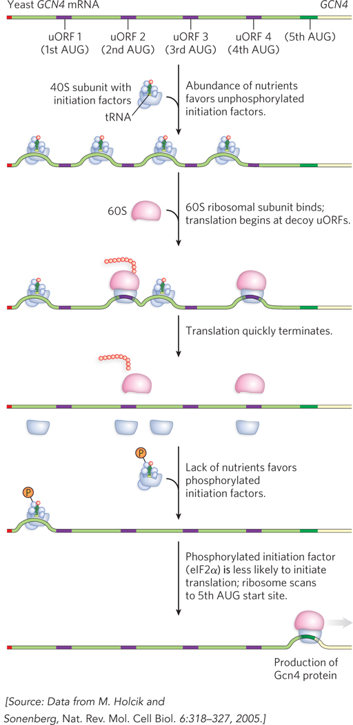
Upstream Open Reading Frames Control the Translation of GCN4 mRNA

Some eukaryotic genes are controlled by short open reading frames located upstream from the gene’s authentic start codon. These upstream open reading frames (uORFs) do not produce functional protein. Instead, they are a gene regulatory mechanism that generally decreases translation by diverting ribosomes, often making them halt and dissociate before reaching the AUG start codon. It might seem that uORFs, rather than regulating levels of gene expression, would simply repress genes under all conditions. However, the effectiveness of uORFs in terminating ribosome activity is altered by phosphorylation of eIF2α. An instance of this type of regulation is observed for the yeast GCN4 gene (general control nonderepressible), encoding a transcription activator that regulates many other genes.

Although the phosphorylation of eIF2α typically down-regulates translation initiation, in S. cerevisiae, low-level eIF2α phosphorylation in response to amino acid starvation induces expression of the transcription factor Gcn4. Because Gcn4 activates transcription of at least 40 genes encoding amino acid biosynthetic enzymes, its induction alleviates nutrient limitation that could otherwise trigger more extensive eIF2α phosphorylation and more general translational repression.

769

The mechanism of activation involves four short uORFs preceding the Gcn4-coding sequence of the mRNA. Located between 150 and 360 nucleotides upstream from the AUG start codon, the uORFs prevent ribosomes from initiating translation at the GCN4 start site when nutrients are abundant. This occurs because ribosomes initiate efficiently at these “decoy” open reading frames instead of at the true protein-coding start site farther downstream (Figure 22-10). When eIF2α is phosphorylated, however, ribosomes are much less likely to initiate translation in general, and thus they have a greater propensity to continue scanning along a bound mRNA without forming an initiation complex. This circumstance favors initiation at the downstream GCN4 start site, leading to expression of the Gcn4 protein.

Figure 22-10: Translational regulation of GCN4 in yeast by upstream ORFs. Unphosphorylated eIF2, present when nutrients are abundant, leads to frequent translation initiation at upstream ORFs (uORFS) and little initiation at the GCN4 gene (first three steps). When eIF2α is phosphorylated, translation initiation in general, including at the uORFs, is reduced; initiation at GCN4 is now more likely (last two steps).

mRNA Degradation Rates Can Control Translational Efficiency

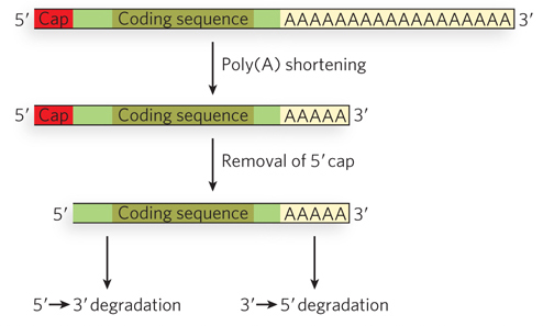
Another important way in which translation is regulated in eukaryotic cells is by the degradation of mRNAs. Translation and degradation of particular mRNAs often show an inverse relationship. Those mRNAs that are efficiently translated tend to be stable in the cytoplasm, whereas those that are poorly translated tend to be degraded more quickly. The discovery of specific pathways of mRNA degradation has led to a better understanding of the close correlation between mRNA translation and turnover. Decay of mRNAs begins with removal of the 3′ poly(A) tail (deadenylation), followed by removal of the 5′ 7-meG cap (Figure 22-11). The mRNAs are then efficiently degraded by exonucleases, some in the 5′→3′ direction, others in the 3′→5′ direction.

Figure 22-11: Eukaryotic mRNA decay. The initial steps involve removal of first the 3′ poly(A) tail then the 5′ cap. Exonuclease-catalyzed degradation of the remaining RNA is then rapid.

770

Removal of the 5′ cap involves the formation of macromolecular assemblies of translationally repressed mRNAs bound to decapping enzymes. Known as processing bodies (P bodies) (see Figure 16-26), these large cytoplasmic structures are easily seen by light microscopy in cells that have been starved or otherwise stressed to induce general translational repression.

Studies of the composition and formation of P bodies suggest that the rates of mRNA translation and degradation are influenced by the relative stability of the mRNA bound in polyribosomes and in P bodies. Regulatory proteins that bind sequences or structures within related groups of mRNAs serve either to recruit those mRNAs to P bodies or to stabilize their interaction with ribosomes. This is an exciting area of active research. P bodies are part of the conserved translational control machinery in eukaryotic cells, influencing patterns of gene expression in cells as diverse as oocytes and neurons.

SECTION 22.2 SUMMARY

  • Eukaryotes have at least three mechanisms of translational regulation: phosphorylation-dependent repression of initiation factors, direct binding of mRNAs by translational repressors, and disruption of the required interaction between eIF4E and eIF4G.

  • Reversible phosphorylation of initiation factors plays a central role in regulating translation initiation in response to cellular conditions. When the eIF2α subunit is phosphorylated, eIF2 forms a stable complex with eIF2B, making these factors unavailable for further rounds of translation initiation.

  • The CPE-binding protein binds sequences in the 3′UTR of maternal mRNAs and contributes both to suppressing translation (in conjunction with Maskin, a 4E-BP-like protein) and to activating translation (through interaction with CPSF).

  • All PUF proteins have characteristic sequence and structural features that enable their binding to the 3′UTRs of mRNAs. Binding leads to translational repression and/or degradation of the transcript, reducing expression levels.

  • In yeast cells, upstream open reading frames (uORFs) in the GCN4 mRNA, preceding the Gcn4-coding sequence, act as ribosome decoys. They prevent ribosomes from initiating translation at the GCN4 start site when nutrients are abundant, allowing ribosomes to initiate efficiently at the uORFs instead of at the authentic start site farther downstream.

  • Efficiently translated mRNAs tend to be stable in the cytoplasm; those that are poorly translated tend to be rapidly degraded. The decay of an mRNA involves removal of the 3′ poly(A) tail, decapping of the 5′ end, then exonuclease-catalyzed degradation.

  • Cytoplasmic foci, known as P bodies in yeast, are sites of mRNA decapping and degradation.